CT in blunt chest trauma: indications and limitations.
Van Hise ML, Primack SL, Israel RS, Muller NL
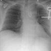
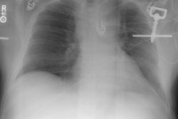
Computed tomography (CT) is the imaging modality of choice in the assessment
of patients with clinical or radiographic findings suggestive of aortic
injury, bone fracture, or diaphragmatic tear following blunt chest trauma.
Contrast material-enhanced spiral CT allows detection of both subtle and
more obvious aortic tears. CT has overall greater sensitivity than radiography
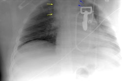
in the detection of pulmonary lacerations and pneumothoraces. CT may be
indicated in cases of suspected tracheobronchial injury. CT is of limited
use in the assessment of rib fractures because such injuries are of limited
clinical significance and can usually be identified at radiography; however,
CT provides optimal visualization of thoracic spine fractures and superior
assessment of suspected sternal fractures or sternoclavicular dislocation.
Targeted spiral CT with sagittal and coronal reformatted images has increased
sensitivity and specificity over that provided by conventional axial CT
in the detection of diaphragmatic injury. Optimal CT assessment requires
careful attention to technique, including the use of intravenously administered
contrast material and multiplanar reconstructed images, as well as an awareness
of potential pitfalls. Although in many cases diagnosis can be made with
confidence on the basis of CT findings, further investigation is often
needed to confirm the diagnosis.