Radiographics 1997 Jan;17(1):27-45. Overview of traumatic injury of the thoracic aorta.
Creasy JD, Chiles C, Routh WD, Dyer RB
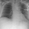
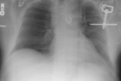
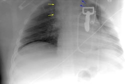
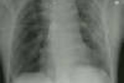
Traumatic injury of the thoracic aorta is a major clinical concern in patients who sustain deceleration or crush injuries. Several mechanical factors may explain the typical locations of thoracic aortic rupture (aortic isthmus, ascending aorta). Understanding these factors and the pathophysiology involved helps the radiologist to recognize aortic trauma at various imaging examinations. Chest radiography is the initial screening examination, and radiographs are evaluated specifically for signs of mediastinal hematoma, an indication of significant thoracic trauma. The most important of these signs include loss of aortic contour, tracheal deviation, ratio of mediastinal width to chest width, deviation of a nasogastric tube to the right of the T-4 spinous process, and depression of the left main-stem bronchus (> 40 degrees below the horizontal). Computed tomography (CT) is used increasingly when results of chest radiography are equivocal. CT can clearly demonstrate mediastinal hematoma, but this finding is also mimicked by several entities, including atelectatic lung, thymus, and pericardial recesses. Aortography is the standard for diagnosis. Traumatic aortic injury is treated urgently with surgical repair. The rare patient who survives aortic injury without surgery may develop a chronic pseudoaneurysm.
PMID: 9017797, MUID: 97170268