Radiographic and CT findings of blunt chest trauma: aortic injuries and looking beyond them.
Kuhlman JE, Pozniak MA, Collins J, Knisely BL
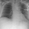
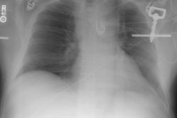
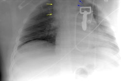
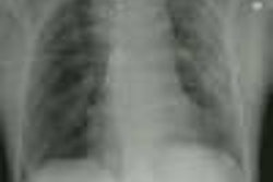
Increasingly, helical CT is being used to screen trauma patients for aortic injury. Most aortic injuries visible at CT occur at or near the level of the ligamentum arteriosus; these injuries manifest as mediastinal hematoma, aortic contour deformity, intimal flaps, intraluminal debris, pseudoaneurysm, and pseudocoarctation. In the process of searching for aortic injury, however, the radiologist should not overlook other serious and more common thoracic injuries. Tracheobronchial tears appear at CT and radiography with persistent pneumothorax, subcutaneous emphysema, "fallen lung" sign, and malposition of endotracheal tube. The ruptured diaphragm, which tears more often on the left, appears asymmetric, irregular, or discontinuous, with herniation of bowel or viscera into the chest. In esophageal rupture, CT and radiography demonstrate left pneumothorax, pneumomediastinum, subcutaneous emphysema, and pleural effusion and atelectasis on the left. CT is better than trauma radiography for depicting fractures of the thoracic vertebral bodies and ribs, as well as for revealing pulmonary contusions and lacerations. CT is also useful for demonstrating unsuspected injuries caused by seat belts. Observation of these injuries should prompt a search for other serious internal organ injuries.