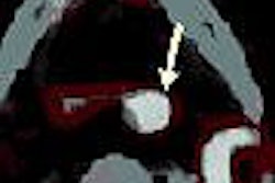
ORLANDO - Scintimammography using a dedicated breast gamma camera shows "high sensitivity and moderate specificity" in breast cancer detection, according to research presented this week at the American Roentgen Ray Society (ARRS) annual meeting.
The research -- to determine sensitivity and specificity of scintimammography for the detection of breast cancer, using pathology as the reference standard -- also noted that scintimammography discovered six occult cancers not found with clinical examination or other imaging modalities. The results were presented by Dr. Angelique Floerke of George Washington University in Washington, DC.
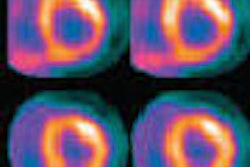
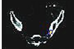
The advantage of scintimammography is "its physiological approach to breast imaging and its ability to interpret images in breast density," Floerke said. "It is well-tolerated by the patient and radiologists, as only four to 10 images are generated for interpretation."
The retrospective study included 146 women who underwent scintimammography with a dedicated breast-specific gamma imaging (BSGI) system developed by Dilon Technologies of Newport News, VA (One of the authors of the study, Dr. Rachel Brem of George Washington University, has a financial interest in Dilon).
Patients were imaged in craniocaudal (CC) and mediolateral oblique (MLO) projections. Patients received an intravenous injection of 30 mCi of Tc-99m sestamibi.
Of the 146 patients, 167 lesions underwent biopsy, which demonstrated 83 malignant lesions. Of those 83 lesions, 16 were ductal carcinoma in situ (DCIS), and 67 invasive cancers were found.
"Of the 83 malignancies -- invasive or in situ -- (scintimammography) correctly identified 80 as malignant, or 94%," noted Floerke. "If we consider just the invasive cancers, 65 of 67 were correctly identified by (scintimammography), resulting in a sensitivity of 97%."
The study reported that scintimammography also identified in situ malignancy in 15 or 16 cases, which yields a sensitivity of 94%. Of the lesion sizes, the median size of invasive cancers detected was 15 mm. The median size of detected DCIS was 7 mm.
Of 114 lesions with a positive scintimammography exam, 80 were invasive cancer, producing a positive predictive value of 70%. Of 53 lesions with a negative exam for malignancy, 50 had no evidence of malignancy on biopsy, yielding a negative predictive value of 94%.
Of the 84 nonmalignant lesions, 82 were benign and two were high risk (one atypical lobular hyperplasia and one lobular carcinoma in situ).
In addition, there were 34 false-positive lesions. "The most common pathology among these was fibrocystic change," Floerke said. "Interestingly, eight patents with a false-positive exam had a biopsy in the preceding two months within the area of increased radiotracer uptake."
Three cancers were undetected by scintimammography -- one was a high-grade DCIS of 10 mm. The lesion was detected at mammography.
By Wayne Forrest
AuntMinnie.com staff writer
May 9, 2007
Related Reading
Solid-state dual-head camera supercharges molecular breast imaging, December 19, 2006
Whole-body FDG-PET of little use in breast cancer staging, November 26, 2006
Preop FDG-PET spots axillary involvement in breast cancer, may obviate biopsy, August 21, 2006
Breast cancer-involved sentinel nodes often contain micrometastases, August 15, 2006
SLN procedure in pregnant breast cancer patients leads to negligible fetal dose, August 14, 2006
Copyright © 2007 AuntMinnie.com