VIENNA - When elbows are injured, the radial head-capitellum view makes a fine adjunct to the standard anterioposterior (AP) and lateral views when more information is needed. But the technique shouldn't be allowed to elbow its way into the standard imaging protocol, according to a presentation Tuesday at the European Congress of Radiology (ECR).
Most radial-head fractures are clearly visualized on AP and lateral views, but additional projections are sometimes necessary, said Dimitris Koumarianos, a radiologic technologist from ELPIS General Hospital of Athens.
The radial head-capitellum view, with angulation of the elbow 45 degrees toward the shoulder, "displaces without superimposition the radial head, the capitellum and the coronal process," he said. The projection was supported in a study by Greenspan and Norman, and recommended as a routine technique for elbow trauma injury (American Journal of Roentgenology, June 1982, Vol. 138:6, pp. 1,186-1,188).
"We undertook this study to check the diagnostic accuracy of this projection," Koumarianos said.
Koumarianos and colleagues retrospectively analyzed the images of 91 consecutive patients presenting with acute elbow trauma. Each patient's x-ray exam included an AP lateral and a radial head-capitellum view. Two radiologists read each set of images twice, analyzing and recording whether the injury could be seen on each view separately, or only one view.
The results showed 31 normal studies. Eleven patients had no evidence of bone injury but positive fat pad signs, and 12 studies showed some bone injury. The majority of fractures were in the radial head and neck (13/20, 65%). There were also two fractures in the coronoid (10%), one in the capitellum (5%), two in the olecranon (10%), and two supracondylar fractures (10%).
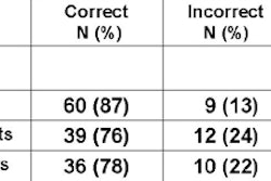
Twelve of 13 fractures of the radial head and neck were seen only in the AP and lateral views. On the radial head-capitellum view, however, only five of the 13 fractures were visualized. There were no significant differences between the techniques in the other fractures.
"For the 13 radial head fractures, the radial head-capitellum view offered no advantage in 10 cases, but it did offer an advantage in three cases," he said. "In half of the cases of the coronoid, (radial head-capitellum) was useful."
Overall, the AP and lateral views demonstrated 95% of all fractures, he said. The radial head-capitellum view failed to demonstrate 40% of all fractures, he said. Radial-head offered an advantage in 30% of all fractures.
Omitting radial head-capitellum would have led to the miss of one fracture: a short intra-articular nondisplaced fracture of the radial had. Still, on the AP and lateral views of this case, both the anterior and posterior fat pads were positive, Koumarianos said.
The group's results are mostly in line with other studies, he said. Grundy et al found that the radial-head capitellum view added information in only 21% of 28 cases, and concluded that it offered no objective benefits in the primary diagnostic rate and treatment for elbow injuries, Koumarianos said (British Journal of Radiology, October 1985, Vol. 58:694, pp. 965-967).
Hall-Craggs and colleagues reported that radial head-capitellum detected only 50% of the elbow injuries, with only one injury that wasn't seen on the standard AP/lateral views (AJR, September 1985, Vol. 145:3, pp. 607-609).
However, a large series by Manns and Lee found that the radial head-capitellum view showed 87% of fractures, with a single fracture not seen on the AP/lateral views, according to Koumarianos (Clinical Radiology, December 1990, Vol. 42:6, pp. 433-436).
Limitations of the Athens study included its small size and the lack of proof that the correct diagnosis was made, Koumarianos said.
"This study suggests that the radial head-capitellum view should be considered a supplementary rather than a standard view for trauma of the elbow," Koumarianos concluded. "Routine use of the radial head-capitellum projection is not justified in all cases of elbow trauma. The recognition of the positive fat-pad sign would be more beneficial than adding a new view to routine elbow trauma radiography. The radial-head view could offer considerable assistance on selected cases only."
By Eric Barnes
AuntMinnie.com staff writer
March 9, 2005
Related Reading
The bends and flexures of forearm and elbow x-ray positioning, November 21, 2002
Copyright © 2005 AuntMinnie.com