A preprogrammed protocol can significantly improve the efficiency of breast ultrasound survey scans compared to standard scanning routines, according to research from Northwestern University Feinberg School of Medicine in Chicago.
"We found the protocol enabled the operator to focus on scanning and findings instead of annotating," said Dr. Holly Marshall. "And the protocol promoted consistency in acquiring images for interpretation."
She presented the team's findings during a scientific session at the 2009 American Roentgen Ray Society (ARRS) meeting in Boston.
Based on its success in improving efficiency in obstetric and hepatic sonography, scanning with preprogrammed protocols has recently been introduced for breast survey scanning, Marshall said. In addition, results from the American College of Radiology Imaging Network (ACRIN) 6666 screening ultrasound trial have led to heightened interest in survey scanning for screening and for evaluating the extent of disease in cancer patients, she said.
"Workflow in the electronic setting is a current issue in many breast centers, as time-saving measures are needed to accommodate increased patient demand and volumes, particularly as many diagnostic evaluations have become multimodality," she said.
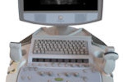
To investigate the usefulness of a preprogrammed protocol for breast ultrasound survey scanning, the Northwestern researchers studied a scanning template installed following a software upgrade to an iU22 ultrasound scanner (Philips Healthcare, Andover, MA).
The protocol uses a template of user-selected annotated views that follow in automated sequence. Once a view is captured, the next preannotated view will appear until the examination is completed, Marshall said. Preannotated views are programmed separately for each breast, as well as for bilateral studies, she said.
The protocol is paused for diagnostic evaluation and image capture of a finding, allowing for the usual lesion characterization techniques to be applied, for orthogonal views to be recorded, and for measurements to be taken, she said.
The researchers compared the technique with routine ultrasound scanning, in which each view was annotated manually by the operator. Thirty women were selected for or requested survey scanning; the group included those with single or multiple palpable masses, dense breasts with higher intermediate risk of breast cancer, mammographic findings, and breast pain.
Each patient was scanned twice using the iU22 scanner with either a L17-5 or L12-5 linear-array transducer, once using the preprogrammed protocol and once using routine techniques. The scanning order was randomized, and survey scans were performed by a breast-certified sonographer with 16 years of experience.
The exams included 11 unilateral exams and 19 bilateral exams. The automated protocol had an average scanning time of seven minutes, a time savings of one minute, 40 seconds, compared with the average routine scanning time of eight minutes, 40 seconds.
For unilateral exams performed using the protocol, the time to complete the study ranged from two minutes, 30 seconds, to 10 minutes. Exams performed using routine techniques ranged from three minutes, 38 seconds, to 10 minutes. In two patients, the protocol scan time was equal to or longer than the routine exam.
Bilateral studies performed using the protocol ranged from five minutes, 14 seconds, to 35 minutes, compared with a range of five minutes, four seconds, to 37 minutes for routine bilateral scanning.
In 26 of the 30 patients, the protocol scan was interrupted, which lengthened scan time. In 13 cases, the study was paused once. The exam was paused twice in six cases, three times in three cases, four times in two cases, and five times in one, according to the study team. In addition, eight pauses for multiple benign masses occurred in one case.
If the protocol was employed on 15 patients a day, the average 25-minute time savings could allow for one extra patient to be scanned per day, five additional patients per week, and 20 additional patients per month, Marshall said.
"Workflow efficiency is promoted by the protocol," she said.
In the future, the researchers believe additional protocols could be designed to further streamline workflow, such as in palpable areas of concern and recording of ultrasound-guided procedures, she said.
By Erik L. Ridley
AuntMinnie.com staff writer
June 3, 2009
Related Reading
Follow-up ultrasound aids in evaluating new lesions from breast MR, April 1, 2009
Automated breast US unit not ready to replace handheld, study finds, March 16, 2009
Ultrasound, MRI perform well in dense breasts, March 8, 2009
3D Doppler evaluation helps identify malignant breast lesions, November 6, 2008
Breast ultrasound CAD helps doctors find smaller lesions, October 10, 2008
Copyright © 2009 AuntMinnie.com