Femoroacetabular impingement (FAI) is one of the causes of premature osteoarthritis in the hip, and occurs when there is a conflict between the proximal femur and the acetabular rim. MR arthrography is particularly useful for the presurgical assessment of existing damage within the joint, according to radiologists and orthopedists in Switzerland.
"The identification of FAI as a cause of cartilage damage and labral tears may allow surgeons to correct it early in its natural history and delay or prevent end-stage osteoarthritis," wrote Dr. Christian Pfirrmann and colleagues from the University Hospital Balgrist in Zurich (Radiology, July 20, 2006).
More specifically, MR arthrography can help differentiate between cam FAI and pincer FAI based on the predominance of either a femoral or acetabular abnormality, they added.
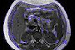
For this study, 50 consecutive patients underwent contrast-enhanced MR arthrographic exams on a 1.5-tesla unit (Symphony, Siemens Medical Solutions, Erlangen, Germany). The sequences included coronal T1-weighted spin-echo imaging, coronal intermediate-weighted fast spin-echo with fat saturation, and sagittal T1-weighted spin-echo. Pfirrmann and co-author Dr. Bernard Mengiardi reviewed the images in consensus and without any knowledge of the surgical diagnosis.
Using a method described in an earlier study, the group measured the nonspherical shape of the femoral head-neck junction in eight positions around the femoral head and neck, with radially reformatted images, to calculate the alpha angle (Journal of Bone and Joint Surgery British, May 2002, Vol. 84:4, pp. 556-560).
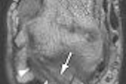
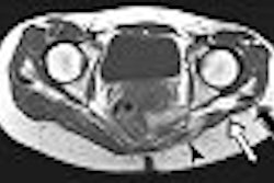
Acetabular cartilage and acetabular labral abnormalities, as well as acetabular bony contours were qualitatively assessed in six positions. The format of an osseous bump at the femoral neck was qualitatively rated as present or absent.
Thirty-three patients were classified as having cam FAI and 17 had pincer FAI at surgery. Notable MR arthrography features of cam FAI were a larger alpha angle (68°) at the anterior position and anterosuperior position (81°). In the pincer FAI, the alpha angle was 54° in the anterior position and 66° in the anteriosuperior position. Also, the acetabulum was significantly deeper in the pincer FAI group (4.8 mm mean depth) than in the cam FAI group (0.7 mm mean depth).
Other MR arthrography features that the group found:
- Larger cam FAI cartilage lesions at the anterosuperior and superior positions
- Larger pincer FAI cartilage lesions at the posteroinferior and posterior positions
- More pronounced labral lesions at the posteroinferior and posterior positions in pincer FAI
- No significant difference in distribution of acetabular osseous abnormalities
- Osseous bump formation more common in cam FAI
The authors acknowledged that the lack of a control group was a weak point in their study. Nevertheless, they emphasized that MR arthrography was adept at the detection and staging of acetabular labrum lesions, which is an important presurgical element. They explained that surgical treatment may be suitable only for patients without advanced degenerative changes.
By Shalmali Pal
AuntMinnie.com staff writer
August 22, 2006
Related Reading
MRI of gluteal contracture backs up physical exam results, July 28, 2006
MR arthrography proves superior to standard MR in labral tears, January 24, 2006
X-ray planning improves hip replacement surgical outcomes, January 18, 2006
MR clarifies paralabral cysts as common source of hip pain, September 6, 2004
Copyright © 2006 AuntMinnie.com