Contracture of the gluteus maximus muscle can be caused by a congenital disorder or intramuscular injection myopathy. Most commonly affecting children and young adults, the condition is diagnosed clinically with a squatting test. Although imaging is not routinely used in gluteal contracture, MRI can help establish diagnosis and evaluate lesion severity.
The physical abnormalities and deformities that result from gluteal contracture include limitation of movement in the affected hip, explained Dr. Clement Chen and colleagues in the American Journal of Roentgenology, in which they outlined the MRI features of gluteal contracture. Chen is from Kaohsiung Veterans General Hospital in Kaohsiung, Taiwan. His co-authors are from other hospitals in Kaohsiung and Taipei.
For this retrospective study, the group looked at MR exams performed on 21 patients (mean age of 22), all of whom had a history of receiving injections in the buttocks. MRI of the pelvis, buttocks, and hips was done on a 1.5-tesla scanner (Signa, GE Healthcare, Chalfont St. Giles, U.K.). The imaging protocol included spin-echo T1-weighted axial images, fat-saturated fast spin-echo proton density-weighted axial images, and spin-echo T1-weighted coronal images. MRI tests were also done on 10 age-matched control subjects.
"On the MRI of the healthy control group, (the gluteus maximus) muscle was symmetric in size, shape, and signal intensity on both sides," Chen's group wrote (AJR online, August 2006, Vol. 187, pp. W169-W174).
 |
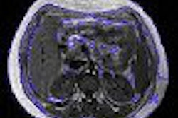
| A 23-year-old woman suffering from bilateral gluteal contracture that is more severe on left side. Patient related history of repeated injections in both buttocks since adolescence. Spin-echo T1-weighted axial image (TR/TE, 566/16) shows atrophic and medial retraction of left gluteus maximus muscle (arrowhead), thickening and retraction of fibrotic cord and distal tendon (white arrow), and displacement of iliotibial tract (black arrow). |
Among the 21 patients, 17 had a contracture of the bilateral gluteus maximus muscles while four had lesions on the right buttock only. All buttocks showed primary features of muscle atrophy and intramuscular fibrotic cord contiguous to the iliotibial track, which manifested as low signal intensity.
"Retraction of the thickened tendon and fibrotic cord accounted for the secondary MRI features," they wrote, such as medial retraction of the affected muscle and a depressed groove at the muscle tendon junction. "The muscle atrophy affected most commonly the upper third of the gluteus maximus, which is related to the injection technique."
 |
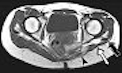
| Same patient as above. Spin-echo T1-weighted axial image (566/16) inferior in relation to image above discloses atrophic left gluteus maximus muscle (arrowhead), thickening and retraction of distal tendon and iliotibial tract (black arrows) posterior to left femur. Angle formed by lines of connecting ischia (black line) and axis of femoral necks (double-headed arrows) measures 9° on right side and 36° on left side. Exaggerated anteverted axis of left femoral neck suggests external rotation of left proximal femur. Chen CKH, Yeh L, Chang W, Pan H, and Yang C, "MRI Diagnosis of Contracture of the Gluteus Maximus Muscle," (AJR 2006; 187:W169-W174). |
The group concluded that the severity of the clinical abnormality can be judged by the presence of thickening and retraction of the fibrotic cord on MRI. In more advanced cases, a depressed groove will appear at the muscle-tendon junction, they added.
In this review, the authors pointed out frontal radiographs of the pelvis were of no use for diagnosing gluteal contracture. Future studies will have to correlate the degree of clinical abnormality with the degree of abnormality on MRI, they stated.
By Shalmali Pal
AuntMinnie.com staff writer
July 28, 2006
Related Reading
MR arthrography proves superior to standard MR in labral tears, January 24, 2006
MR clarifies paralabral cysts as common source of hip pain, September 6, 2004
Ultrasound confirms snapping hip, August 21, 2002
Copyright © 2006 AuntMinnie.com