SAN FRANCISCO - MRI can answer many of the major questions surrounding term infants who may have sustained perinatal brain injuries, according to a presentation this week at the Pediatric Academic Societies (PAS) meeting.
Dr. Mary Rutherford from Hammersmith Hospital in London discussed when and why MRI should be used in this patient population: for ascertaining the causes and symptoms of possible brain injury, for making a specific diagnosis, and for guiding clinical management.
However, Rutherford stressed that MRI is not a substitute for cranial ultrasound, which she recommended for any infant with seizures. When the ultrasound results are abnormal, MRI can offer more detail, especially in the neonatal period.
In her talk, Rutherford first reviewed when MRI should be performed. Perinatal brain injury is the most common indication for neonatal MRI, she said. In the event of trauma, MRI should be used as part of the intervention protocol as soon as possible. The modality also has value if a decision needs to be made as to whether the baby can breathe on his or her own, and can be moved out of intensive care.
MRI can also be used to help predict outcomes: imaging done two to three weeks after birth can reveal mature brain lesions, Rutherford said. Finally, postmortem MRI can offer a greater understanding of brain development.
Next, she discussed how MRI should be done. In terms of equipment, Rutherford suggested that a small coil be used. A still infant (secured in either a transporter or vacuum papoose) must either be asleep or sedated. Imaging must be done in at least two planes with at least two sequences, she said, adding that these are not necessarily the same sequences used for adult neuroradiology.
In answering an audience member question, Rutherford said that her group does not find proton density-weighted MR to be of any added value and that they rely more on T1- and T2-weighted imaging. Also, they do not routinely use contrast media.
In addition, an experienced clinical team must be willing to dedicate the required time for an MR exam and be able to handle any unexpected situations (e.g., baby takes awhile to settle, baby wakes up before the end of the exam), she said.
Clinicians should also be familiar with the normal imaging appearance of the neonatal brain and the various abnormalities that can occur during brain development, Rutherford said. The radiologist who reads the images should be well-versed in the imaging appearance of the brain on different MRI sequences, as this can offer useful information on the timing of the injury or insult, she added. For example, some myelin is visible starting at 37 weeks, while hematomas can change over time.
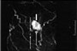
Rutherford stressed that clinicians need to supply the radiologist with as much clinical information as possible on etiology, fetal distress, Apgar scores, gestation, and resuscitation. For instance, a baby with seizures may have a normal Apgar score. Signs that the radiologist will want to look for on the MRI include white-matter infarction, hemorrhage, infection, and hypoglycemia, which will appear as parasagittal infarction.
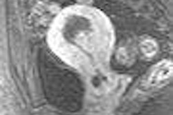
MRI can also help clarify results in cases of hypoxic-ischemic encephalopathy (HIE), which is associated with widespread white-matter involvement and cognitive impairment, although not always motor impairment. In a study of 270 HIE patients conducted at her institution, Rutherford said that 48 patients had an acute sentinel event and HIE. In that group, 63% had basal ganglia and thalamic lesions, which are associated with cerebral palsy, she added.
Finally, Rutherford summarized how MRI can help clinicians with newborn patients during the first few weeks of their lives by offering an appreciation of the evolution of lesions, as well as lesion detection and evaluation. MRI is also an important component in addressing the medicolegal aspects that are often associated with neonatal brain injury cases, she said.
By Shalmali Pal
AuntMinnie.com staff writer
May 3, 2006
Related Reading
Study shows premature babies can feel pain, April 5, 2006
Infant with hypoplastic left heart syndrome successfully treated in utero, January 31, 2006
Preop MRI to measure CBF may benefit neonates with heart defects, December 29, 2004
Neonatal MRI predicts cerebral palsy in neonates at risk, October 15, 2004
Copyright © 2006 AuntMinnie.com