A study based at the Cleveland Clinic in Ohio shows surgical outcome in refractory epilepsy can be predicted by PET imaging, even in patients with frequent seizures or spiking activity.
Dr. Roland Talanow of the Cleveland Clinic presented the research results at last month's RSNA 2007 meeting in Chicago. PET imaging is performed for the presurgical workup of patients with refractory seizures despite medical treatment, he noted. However, some patients present with seizures (ictal) or frequent spikes (abnormal electroencephalogram [EEG]) during a PET scan. To date, it has not been known definitively how the PET results correlate with surgical outcomes in patients with frequent spiking activity on an EEG and ictal activity shortly after injection of FDG.
In the study, 294 patients with refractory epilepsy were identified and underwent PET imaging with EEGs for presurgical workup. The analysis included 143 females and 153 males, ranging in age from one day to 64 years. Patients fasted for at least four hours prior to the exam and were seizure-free at least 24 hours before the study.
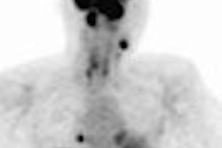
A dose of 5-15 mCi of FDG was injected intravenously for adults. Pediatric patient dose was adjusted accordingly based on weight. Scanning was performed on an ECAT HR+ PET scanner (Siemens Medical Solutions, Malvern, PA).
The EEG, which monitored activity from 30 minutes before to 30 to 45 minutes after injection of FDG, was classified as true interictal (normal), interictal with frequent spikes, and ictal (clinical seizure present). The metabolism level on the PET scan was also defined as hypermetabolic, normal, and hypometabolic.
All patients underwent surgical treatment for seizure focus removal, and surgical outcome was segmented intro three categories: seizure-free, improved, and failed.
In the analysis, researchers found that 260 (88%) of the 294 patients had true interictal PET imaging, while 14 (5%) had ictal imaging. In addition, 20 cases (7%) had interictal frequent spiking activity based on EEG results.
In analyzing the groups more specifically, the researchers determined that among the 260 patients with true interictal PET, 95% of the cases demonstrated hypometabolism in the area of resection, while 85% were seizure-free or improved, and 5% of the patients displayed normal metabolism, with 83% showing no signs of seizure.
Of the cases with interictal but frequent spiking activity, nine (45%) of the patients demonstrated hypermetabolism, another nine (45%) showed hypometabolism, and two (10%) had normal metabolism in the resected regions. In the group with ictal PET, six (43%) of the patients showed hypermetabolism and eight (57%) had hypometabolism in the resected regions.
In addition, the seizure-free rate was 92% in patients with frequent interictal spikes or ictal EEG with PET findings of hypermetabolism. Among the patients with frequent spikes or ictal EEG with PET findings of hypometabolism, the seizure-free rate was 100%. With a normal EEG, the seizure-free rate also was 100%.
"We have investigated all three patient populations -- seizure-free, with frequent spikes, and with clinical seizures -- and have shown that there is a good correlation between frequent spiking activity and ictal EEG and hypermetabolism," Talanow said. The researchers also concluded that "PET hypermetabolism and hypometabolism in patients with frequent spiking activity and ictal state well correlated with surgical outcome."
By Wayne Forrest
AuntMinnie.com staff writer
December 13, 2007
Related Reading
Seizure assessment guidelines unveiled, November 21, 2007
PET changes oncology management, study shows, July 19, 2007
PET/CT moves closer to diagnostic standard of care, March 1, 2007
PET reveals limbic dysfunction in mesial temporal lobe epilepsy, December 19, 2006
Drawing-induced epilepsy points to frontoparietal lobe involvement, March 24, 2006
Copyright © 2007 AuntMinnie.com