VIENNA - The use of dual-modality PET/CT for tumor staging (T), lymph node metastasis staging (N), and distant metastasis staging (M) is more accurate than CT, PET, or side-by-side PET and CT, according to research presented at the European Congress of Radiology (ECR) on Monday. In addition, the use of PET/CT for oncological staging had a significant impact on patient therapy management when compared with the other modalities.
"Stage adapted therapy improves patient survival in a wide variety of malignant tumors," said Dr. Patrick Veit, a research fellow from the department of diagnostic, interventional, and neuroradiology at the University Hospital of Essen in Essen, Germany.
"Tumors are often widely staged with CT, which provides morphological and anatomic data, but it has limited functional assessment," he said. "On the other hand, PET delivers the functional data but is limited in the anatomical assessment. The PET/CT scanners combine the advantages of both modalities."
Veit and his group examined 260 different patients (167 female and 97 male), and conducted primary tumor staging on 112 patients and suspected recurrent disease staging on the remaining 148 patients. They examined various malignancies: 57 lung cancers, 47 head and neck cancers, 44 gastrointestinal tract cancers, 23 liver cancers, 22 thyroid cancers, 21 metastases of unknown primary tumors (CUP syndrome), and 18 genitourinary system cancers.
The patients were imaged on a Siemens Medical Solutions' (Erlangen, Germany) Biograph PET/CT system with a field-of-view of head to upper thighs. Oral and intravenous contrast agents were used for the CT data, which is the standard protocol for the department, according to Veit. Each patient was injected with 350 MBq 18F-FDG one hour prior to scanning, and CT-based PET attenuation scanning was performed.
For the study, all images came from the same procedure and were broken out to their individual components (PET, CT, PET plus CT) for investigative purposes in comparison with the fused PET/CT images, noted Veit.
The PET images were evaluated by two nuclear medicine specialists who performed both qualitative and quantitative assessments on the data. The qualitative assessment was performed on the basis of increased glucose metabolism, while the quantitative assessment was conducted on the basis of standardized uptake values (SUV).
"An SUV of 2.5, extrahepatic, and an SUV of 3.5, intrahepatic, were considered to be malignant," Veit said.
The CT images were evaluated by two radiologists in consensus and were performed on the basis of lymph-node size assessment. The PET plus CT images were reviewed by a nuclear medicine specialist and a radiologist, in consensus, on two side-by-side monitors, one for each modality.
The images were evaluated on the basis of increased glucose metabolism, which was considered pathological, and for lymph node assessment, which was conducted independent of size. The PET/CT fused images were evaluated for the same conditions as the PET plus CT, and it was also conducted by a nuclear medicine specialist and a radiologist in consensus, Veit noted.
"The image evaluation was conducted, of course, for detection of primary or recurrent tumor," he said. "The analysis was done for overall and separate TNM stage and re-evaluated for its impact on patient management."
Histopathology and a clinical exam served as the research team's standard of reference. The group reported a histological reference for the T-stage patients of 77, the N-stage patients of 72, and the M-stage patients of 57. A clinical follow-up was conducted on the entire patient cohort in a mean of 311 days (± 125 days), Veit said.
For the T stage, PET/CT correctly staged 63 patients, with six overstaged and eight understaged patients. PET plus CT correctly staged 55 patients, with 10 overstaged and 12 understaged. CT alone correctly staged 51 patients, with 11 overstaged and 15 understaged. PET alone correctly staged 49 patients, with six overstaged and 22 understaged.
 |
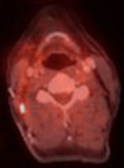
| A 48-year-old male with carcinoma at the base of the tongue. CT and PET images (left and right, respectively) show the pathology. However, the fused PET/CT image indicates osseous infiltration, which was later confirmed by histological pathology. Images courtesy of Dr. Patrick Veit, the University Hospital of Essen, and the ECR. |
For the N stage, PET/CT correctly staged 240 patients, with 12 overstaged and eight understaged patients. PET plus CT correctly staged 230 patients, with 18 overstaged and 12 understaged. PET alone correctly staged 226 patients, with 19 overstaged and 15 understaged. CT alone correctly staged 197 patients, with 27 overstaged and 36 understaged.
For the M stage, PET/CT correctly staged 248 patients, with one overstaged and eight understaged patients. PET plus CT correctly staged 245 patients, with five overstaged and 10 understaged. PET alone correctly staged 231 patients, with one overstaged and 28 understaged. CT alone correctly staged 230 patients, with seven overstaged and 23 understaged.
Of the 260 patients examined by the research team, 218 (84%) were correctly staged with PET/CT, 197 (76%) with PET plus CT, 166 (64%) with PET alone, and 163 (63%) with CT alone, Veit reported
The impact on therapy was significant in all the head-to-head comparisons. In PET/CT versus PET plus CT, 6% (16 of 260) of the patients had an impact on their treatment plan. In the PET/CT versus CT comparison, 15% (39 of 260) of the patients had an impact on their treatment plan, and in the PET/CT versus PET showdown, 17% (43 of 260) of the patients had an impact on their treatment plan, according to the researchers.
On the basis of their research, the team determined that combined PET/CT with its fused images was the superior tool for oncological staging, and that its use has a substantial and significant impact on patient management.
"Combined PET/CT can be recommended as a first-line diagnostic tool for a variety of oncological diseases," Veit stated.
By Jonathan S.
Batchelor
AuntMinnie.com staff writer
March 7, 2005
Related Reading
PET improves on CT in staging esophageal cancer, February 3, 2005
PET and CT combination helps spot recurrent cancer, December 30, 2004
PET/CT improves detection of metastatic colorectal cancer, December 16, 2004
MR, PET/CT show low sensitivity for melanoma, November 30, 2004
Copyright © 2005 AuntMinnie.com