Current ultrasound-based methods for measuring cardiac blood volume are problematic, as the constant motion and deformation of the heart creates a turbulent blood-flow profile, according to Oliver Kripfgans, PhD, who participated in the research. In addition, arterial catheters are typically used for this application. The use of 3D ultrasound could enable more accurate measurements without the use of catheters, he said.
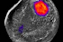
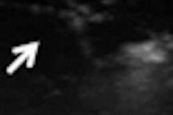
The researchers used a 4D (3D with motion) ultrasound scanner (Vivid 7, GE Healthcare, Chalfont St. Giles, U.K.) to examine a latex tube filled with a fluid that mimicked the properties of blood. Ultrasound scans were performed on the tube as a cardiac bypass pump circulated the fluid at rates of 60 and 80 beats per minute.
Researchers calculated a mean volume flow estimate from C-plane integration and power Doppler-based partial volume correction. They also acquired ultrasound scans with varying numbers of 4D volumes.
They found that ultrasound enabled angle-independent estimates of volume flow to be achieved with less than 10% error when using a minimum of 50 volumes per acquisition. Total imaging time was about five seconds for 50 volumes, which indicates that 4D ultrasound can be used for essentially real-time noninvasive measurement of cardiac output and volume flows.
The researchers are planning further evaluation of the technique in preclinical models and humans, Kripfgans said.
"Setting an ultrasound transducer onto one's chest and accurately reading cardiac output within 30 seconds would be a great improvement," Kripfgans said.