Whole-breast ultrasound tomography has much to offer the world of breast imaging, with resolution approaching that of breast MRI, according to researchers from the Karmanos Cancer Institute at Wayne State University in Detroit.
Not only can it now provide comparable visualization of breast anatomy to MRI, but ultrasound tomography's positive predictive value appears to be a promising improvement over the combination of mammography and conventional ultrasound, said Neb Duric, PhD, an imaging program leader at Karmanos. The researchers also believe that ultrasound tomography shows potential for predicting response to therapy and improving the prediction of breast cancer risk.
Duric presented the institute's clinical experience with breast ultrasound tomography during a session at the American Association of Physicists in Medicine (AAPM) annual meeting in Philadelphia in July.
Although mammography is the gold standard for breast imaging, it suffers from a high biopsy rate of benign lesions and a significant false-negative rate for women with dense breasts, and it's least effective for women at highest risk of cancer, Duric said. It also uses ionizing radiation and is uncomfortable for patients.
Breast MRI is taking an increasingly prominent role, but it's fairly expensive and has a relatively long exam time. Ultrasound, on the other hand, is very cost-effective and performs well in dense breasts. However, the modality is operator-dependent and not well-suited to screening. It also offers relatively low specificity, Duric said.
In an attempt to address the trade-off between the high image quality of MRI versus the low expense of the other modalities, the Karmanos researchers have investigated the use of whole-breast ultrasound tomography technology, he said.
"We want to elevate ultrasound's role in breast imaging, and we want to address this trade-off as a way of finding a middle ground for breast screening diagnosis," Duric said.
In contrast to conventional ultrasound, an ultrasound tomography system surrounds the entire breast with a ring of sensors, allowing for detection of the ultrasound signal scatter field from all angles of transmission, he said. A variety of processing techniques are then used to combine and fuse the images into one image.
To see if an operator-independent whole-breast ultrasound system can offer added value in breast imaging, the Karmanos researchers are investigating its in vivo performance in a clinical setting.
Prototype
The institute's prototype whole-breast ultrasound tomography system, called SoftVue, yields in-plane resolution of 0.5 to 2 mm and out-of-plane resolution of 5 mm. Scan time is approximately one minute.
It has a 20-cm diameter ring, 256 transmitting and receiving elements with 0.5 x 15-mm apertures, 256 data channels, and 2.0-MHz frequency. A full slice is acquired in 30 msec and a full breast scan takes 45 to 60 seconds.
"We typically generate somewhere around 100 slices at 1-mm intervals," he said.
The system has a memory buffer of 22 GB and typically holds 150 slices. Patient throughput is approximately five per hour.
More than five years in development, the system has been used in more than 500 patient exams and has performed more than 100,000 tomograms. Last year, the Karmanos Cancer Institute spun off a company called Delphinus Medical Technologies of Detroit to commercialize the technology.
The researchers are testing their system in three ongoing clinical studies, evaluating its ability to detect breast cancer, to measure therapy response in breast cancer patients undergoing neoadjuvant chemotherapy, and to assess breast cancer risk by using breast density as a modifiable risk factor for breast cancer, Duric said.
US tomography vs. MRI
First, the researchers compared their ultrasound tomography data with MRI, due to its role as an imaging gold standard and because the modality acquires image data in a similar geometric fashion, Duric said.
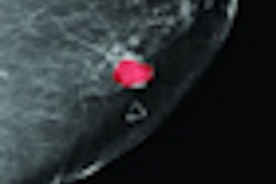
 |
| Breast anatomy shown on breast ultrasound tomography image (left) and breast MRI (right). All images courtesy of Neb Duric, PhD. |
Though its resolution isn't quite as good as MRI, the ultrasound tomography system overall offers similar sensitivity for different tissue types, he said.
"We're pretty confident that the breast architecture that we see is largely real and comparable to what MRI sees," Duric said.
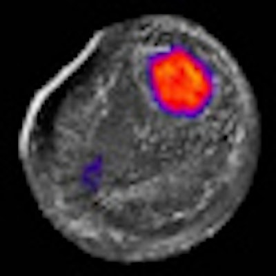
For cancer imaging, pixels are examined in the three sets of ultrasound tomography images (reflection, sound speed, and attenuation). Thresholds are then assigned based on prior evidence showing that cancer tends to be highly stiff and dense; therefore, cancer should have high sound speed and high attenuation.
Only pixels that exceed both thresholds simultaneously are displayed, allowing for the isolation of regions that are biomechanically different, he said.
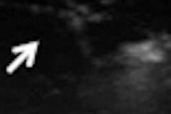
 |
The study team found that dynamic contrast-enhanced MRI and ultrasound tomography turned in similar performance in small tumors, as well as in depicting necrotic regions from large tumors.
To quantify these benefits even further and see how this information could be used for diagnostic purposes, the researchers studied a sample of approximately 100 patients using the latest version of the prototype.
Basic measurements such as average sound speed of tumors and the average attenuation of tumors relative to background tissue were tabulated from the three sets of ultrasound tomography images. A set of predictive functions were created based on these measurements and other factors such as tumor shape.
Based on biopsy results, lesions were plotted as data points on a chart.
 |
Using a data cutpoint that yielded 100% negative predictive value, ultrasound tomography had a positive predictive value of 82% and a specificity of 81%. In comparison, the same process with traditional ultrasound had a 53% positive predictive value and a specificity of 28%.
"This is by no means a definitive clinical trial test, but it does suggest some potential for improvement in generating better [positive predictive value] by using these other properties based on the biomechanical properties of tissue," he said.
Therapy monitoring
The institute is also exploring the technology's benefits when used in monitoring neoadjuvant chemotherapy.
 |
As chemotherapy takes effect over time, the tumor shrinks in size and falls below the pixel visualization threshold.
"More importantly, as far as we're concerned, is that we're also seeing an effect where the average sound speed of the breast is also declining," he said. "And we see this as a somewhat independent marker of the size of the tumor, because what it's telling us is that the tumor is getting softer and softer."
In further analysis of their sample of 11 neoadjuvant chemotherapy monitoring patients, the researchers found a significant difference between patients who responded fully to chemotherapy and those who had a partial response. The average starting sound speed of the responders was lower and the rate of tumor decline was much steeper than for the partial responders, he said.
"So we're thinking now -- it's a working hypothesis as we collect more data -- that the starting point can form a basis of a predictive model to allow us to predict who's going to achieve partial versus complete response," Duric said.
The researchers are also looking at applying their technique for measuring breast density. In-progress research indicates that ultrasound tomography sound speed data are correlating well with percent parenchyma data from MRI, according to Duric.
The researchers acknowledged a number of limitations to their work, including its nonprospective nature and a lack of screening studies.
"We need larger trials, and that's what we're doing in the future," Duric said.
By Erik L. Ridley
AuntMinnie.com staff writer
September 13, 2010
Related Reading
Delphinus raises venture capital, May 26, 2010
Conn. breast density law causes conundrum for mammo sites, April 29, 2010
Automated breast US improves breast cancer detection, March 5, 2010
Karmanos launches breast US spin-off, November 17, 2009
Automated whole-breast US doubles cancer detection, September 10, 2009
Copyright © 2010 AuntMinnie.com