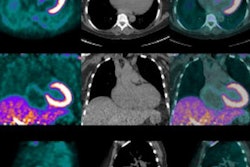
The prospective study included 13 patients with active ankylosing spondylitis who underwent early fluoride-PET scans six minutes after injection of fluoride as part of the PET/MRI protocol. Images were analyzed for diagnostic quality on a four-point scale. The iliac and the sacral part of each sacroiliac joint were subdivided in an upper and a lower part, resulting in four sacroiliac joint quadrants per side. All areas were then evaluated for the presence of bone marrow edema, fatty deposits, sclerosis, ankylosis, and fluoride uptake on blood-pool and mineralization phases of PET.
Mean MR image quality was 3.0 ± 0, while mean image quality for the blood-pool-phase fluoride-PET was only 2.2 ± 0.4. When images from the two modalities were combined, mineralization-phase fluoride-PET/MRI focal uptake was seen in 45 sacroiliac joint quadrants, while blood-pool-phase fluoride-PET/MRI focal uptake was found in only 25 sacroiliac joint quadrants.
On the mineralization phase of fluoride-PET/MRI, sacroiliac joint quadrants showing bone marrow edema alone or a combination of edema with other ankylosing spondylitis lesions had a significantly higher percentage of focal uptake than on blood-pool-phase fluoride-PET/MRI.
Two-phase fluoride-PET/MRI of the sacroiliac joints is feasible, Sawicki and colleagues concluded. However, they had concerns about the efficacy of blood-pool-phase fluoride-PET, saying that it offered no added diagnostic value over standard mineralization-phase fluoride-PET/MRI in patients with active ankylosing spondylitis.