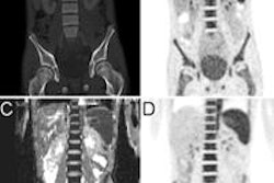
Dr. Julia Fielding and colleagues conducted a pilot study that included five women with cervical cancer (clinical stage greater than IB), who underwent PET/MR of the pelvis before and after initial treatment.
Two radiologists interpreted the MR and PET images; Fielding's group considered smaller tumor size and enhancement of the treated tumor on MR images and decreased standardized uptake values (SUVs) on PET images as indications of response to therapy. Four of the women underwent external-beam radiation therapy and one had surgery.
The women's pretreatment exams showed maximum tumor sizes of 1.7 cm, 1.7 cm, 4.7 cm, 4.8 cm, and 8.7 cm; SUVs were 12.8, 4.1, 4.9, 8.1, and 25.2, respectively.
Both PET and MRI results correlated with clinical staging, leading the researchers to conclude that combined functional and anatomic imaging may be a viable alternative to or confirmation of clinical staging of advanced cervical cancer.