Ultrasonography can easily detect various pulmonary conditions with a high degree of accuracy, despite previous reports to the contrary, according to presenters at the Chest 2006 meeting in Salt Lake City.
"We have been told, even very recently, that the lungs are off-limits to ultrasound," said Dr. Daniel Lichtenstein, director of the medical intensive care unit at the Hospital Ambroise Paré in Boulogne, France. "But lung ultrasonography is really a simple matter," he added, speaking at a symposium on pleural ultrasound at the meeting, which was organized by the American College of Chest Physicians.
Rather than offering just a structural view, ultrasound takes advantage of certain phenomenology in the lungs to produce certain unmistakable images that tell a definitive story of what is happening in the lungs, Lichtenstein said. In addition, using ultrasound could limit the number of CT or x-ray exams that a patient undergoes, thereby reducing the exposure to radiation, he said.
"Using very simple signs, we will be able to describe most of the acute situations," he said. Lichtenstein outlined some of the critical and easily recognizable signs in lung sonography:
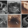
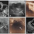
The "Bat" sign (as in the Batman) provides the landmarks for the location of the ribs and the lungs.
The "A" sign appears as a horizontal line across the monitor. These lines occur in a descending pattern equidistant from each other.
The "B" sign has laser-like rays descending from the pleural line. This sign resembles one of the alien-structures that Tom Cruise battled in the movie, "War of the Worlds."
The presence or absence of these signs can aid clinicians in diagnosing thoracentesis, detecting pneumothorax, and distinguishing pulmonary edema from chronic obstructive pulmonary disease, pulmonary embolisms, and other conditions, Lichtenstein said.
In another talk, Dr. Peter Doelken urged his fellow chest specialists to become experts in pulmonary ultrasonography -- something that can be achieved in a very short time, he added. Doelken is an assistant professor of medicine at the Medical University of South Carolina in Charleston and served as the symposium's chairman.
A strength of ultrasound is its ability to detect pleural fluid and even determine the composition of that fluid, Doelken said.
However, ultrasound does have its pitfalls, he stressed, including a misreading of the organs. Doelken advised the first thing a clinician should do when performing a sonographic exam is to find the kidneys and liver, then go on to locate the diaphragm.
In another talk, Dr. Paul Mayo suggested that once pleural effusions are identified, ultrasound can help the clinician gain access to those effusions. Mayo is the director of the medical intensive care unit at Beth Israel Medical Center and a professor of medicine at Albert Einstein College of Medicine, both in New York City.
Mayo laid out three cardinal rules for sonographic identification of pleural effusion:
- Is there an identifiable, echo-free space?
- Have the typical anatomical boundaries been defined and located?
- Are there typical dynamic changes?
The important anatomical boundaries are the diaphragm, the chest wall, and the location of the lungs, he said, adding that the location of the diaphragm can differ among patients. For example, the diaphragm in patients who have undergone coronary bypass graft surgery can be "extraordinarily high," he noted. Mayo said that he has seen serious complications when the clinician misread the position of the diaphragm and pierced the liver, instead of the lungs, to remove fluid.
Neophyte pulmonary ultrasonographers should concentrate on finding "great big black pleural effusions" for at least their first 50 cases, Mayo said. After becoming more comfortable with ultrasound, "you will start to find those gray pleural effusions and, after having done 100 of them, you can chase after the pleural effusions that no one else can see," he added.
By Edward Susman
AuntMinnie.com contributing writer
November 22, 2006
Related Reading
The future of ultrasound: Used by many, understood by few, October 27, 2006
CT lung cancer screening reduces mortality, October 26, 2006
US in the ICU: An essential tool in critical care treatment, February 3, 2006
Copyright © 2006 AuntMinnie.com

