When patients are diagnosed with mild cognitive impairment, it remains unclear if or when they might progress to Alzheimer's disease. But a new study presented Thursday at the American Society of Neuroradiology (ASNR) meeting suggests the transition is inevitable, and that hippocampal volume on MRI roughly predicts how soon it will happen.
Dr. Maciek Bobinski and colleagues from the New York University Medical Center based their conclusions on a series of biannual exams that measured cognitive status and hippocampal volume three times over four years.
According to Bobinski, the study drew upon patients and normal controls (generally spouses of patients) included in a longitudinal database at the university's Center for Brain Health, the neuroimaging core of the NYU-based Aging and Dementia Research Center.
The researchers selected 30 subjects from the database, all of whom had undergone three full diagnostic workups over four years. In addition to medical, neurologic, psychiatric, and neuropsychologic evaluations, patients underwent MR scans using standard diagnostic sequences and a research sequence involving 1.2-mm sagittal T1-weighted sections.
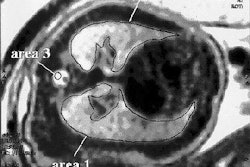
Hippocampal volume was determined using MR landmarks previously validated by post-mortem histology and reported by the researchers (Neuroscience, December 1999, Vol. 95:3, pp. 721-725).
Twenty subjects were patients diagnosed with mild cognitive impairment (MCI) at baseline and first follow-up; the remaining 10 were age- and education-matched controls who remained cognitively normal at the first follow-up. As of the second follow-up, 10 of the MCI patients had developed mild Alzheimer's disease (AD).
The researchers then looked at differences in hippocampal atrophy -- an established hallmark of MCI and AD -- in all three groups. Because the normal hippocampus varies in size relative to a person's height, weight, and sex, the researchers used head size as a standardizing covariant.
It turned out that the MCI patients who converted to AD after four years had 21% less hippocampus than normal controls at baseline, while the non-converting MCI patients had only 10% less. Hippocampal volume at baseline predicted the four-year progression to AD with 85% accuracy, Bobinski said in an interview with AuntMinnie.com.
But all of the MCI patients experienced hippocampal atrophy at similar rates: those who converted to AD lost an average of 3.4% in volume per year compared to 3% per year for the non-converting MCI patients. Those losses contrasted notably with the normal controls, who lost only 1.1% of hippocampal volume per year over four years of aging.
"Once you are clinically labeled as an MCI patient, you have a very high probability of developing Alzheimer's disease, and it's only a matter of time," said Bobinski. "Since right now there is no well-established treatment to stop Alzheimer's disease, it's kind of depressing."
However, Bobinski said, "If you start really aggressive treatment at the very early stage -- not when the patients are diagnosed with Alzheimer's disease, but when they are diagnosed and labeled as MCI -- then you have a higher chance of prolonging this period and hopefully preventing conversion to AD in their lives, since all of them are in their 70s or 80s."
By Tracie L. ThompsonAuntMinnie.com staff writer
June 11, 2004
Related Reading
Frontal cortex tied to implicit memory retention despite dementia, June 10, 2004
Medicare to decide soon on Alzheimer's imaging, June 8, 2004
Do meds to slow dementia really work? MRS may hold the answer, September 12, 2003
Loss of brain volume on MR may precede clinical signs of Alzheimer’s, September 5, 2003
Alzheimer's disease, Part I: PET yields surprising findings in hippocampus, September 7, 2001
PET and MRI race to detect early Alzheimer’s, January 22, 2001
Copyright © 2004 AuntMinnie.com