The use of three-dimensional imaging for medical applications has grown by leaps and bounds in recent years. But even the most advanced 3D-rendered image is still constrained by the limitations of the medium on which it's presented: a 2D computer monitor.
A company called Volume Interactions is hoping to change the 3D visualization paradigm with Dextroscope, a new system designed to make 3D images more intuitive and interactive. The company launched the system in April, and is gearing up for a splash at the upcoming RSNA meeting in Chicago.
The Dextroscope technology is based on stereoscopic imaging, in which a user wears a set of active stereoscopic glasses while viewing 3D images. The configuration creates the impression of a 3D image volume hanging in space in front of the user, who can then make measurements or interact with the volume with various manipulation tools, according to Joseph Balogh, general manager of Volume Interactions USA, the Princeton, NJ-based unit of company, which has its global headquarters in Singapore.
The company's RadioDexter software accepts conventional medical images from multiple modalities, such as MRI, CT, and volumetric ultrasound, and co-registers them. It then processes the information for display on the Dextroscope, a console about the size of an ATM machine.
Rather than mount a display directly in front of the user, the monitor is located above the user, who views a reflection of the (virtual) image in a mirror located directly in front of them while wearing stereoscopic glasses. The stereoscopic effect is created by having the Dextroscope display separate images for each eye, with the images synchronized using infrared emitters in the glasses.
 |
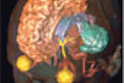
| Dextroscope users wear stereoscopic glasses and view images projected onto a mirror to create the illusion of a volumetric image suspended in space. |
The end result of all this technology is an image that appears to float in space 8-12 inches in front of the user. Balogh believes Dextroscope creates a different viewing experience than what's currently available with 3D images on 2D displays.
"This is fundamentally different than whatever is out there today, which is great 3D imaging, except that it's constrained to a two-dimensional display," he said. "Ours is no longer a two-dimensional display, it's a three-dimensional virtual object."
Dextroscope's console includes a number of tools for working with and manipulating images. The system can track the movements of the user's hands, enabling the user to grab and move the virtual image with the left hand, while the right hand employs a special multipurpose stylus that can perform specialized operations such as cropping, measuring, and calculating angles in space. Because the image is in 3D, users are able to measure image volumes, Balogh said.
|
Note: Video clip may take several minutes to load. Having trouble viewing this clip? Click here to view full size clip or to change format. |
| This video clip demonstrates a Dextroscope user manipulating virtual image volumes in real-time. Images and video courtesy of Volume Interactions. |
Target markets
Volume Interactions sees neurosurgery as the first target market for Dextroscope. While radiologists are used to working with 2D image slices that represent larger volumes, neurosurgeons often haven't developed this ability, Balogh said. Enabling them to plan surgeries in 3D can be an advantage in understanding the patient anatomy they are about to encounter.
"Neurosurgeons operate on three-dimensional patients, so when they go into the OR they are looking down at a 3D object -- the patient's head and brain. With our system they are looking at the imaging of that in the same way: in a 3D stereoscopic view with depth perception," Balogh said "It's much easier for them to get the concept of the anatomy and the relationships of the various complexities of that anatomy and pathology, and then translate that to what they're going to see in the OR."
Cardiologists might also be interested in the system due to its ability to take data from 64-slice CT heart studies and turn it into 3D images, and the system will support dynamic imaging in future releases. Oral and maxillofacial surgeons and otolaryngologists are also potential customers.
Other advantages of Dextroscope include its support for interactivity and its ability to display fused multimodality images. The system is typically connected to a PACS network and accepts DICOM-formatted images, Balogh said.
While radiologists aren't the primary market for Dextroscope, Volume Interactions sees the system as a tool that can facilitate communications among radiologists and multiple surgical specialties. Radiologists and surgeons can sit down at a Dextroscope station together to plan surgeries in what the company believes is a more interactive way than what's possible on workstations with conventional 2D monitors.
Volume Interactions launched the system at the American Association of Neurological Surgeons (AANS) meeting in April, and the company has installed six units in the U.S. and 20-30 worldwide. At the upcoming RSNA meeting, the firm plans to unveil DextroBeam, a version of the system designed for education and group conferencing that would use dual projectors to display 3D images in locations such as a large meeting hall.
As Volume Interactions moves Dextroscope to market, it will have some deep pockets behind it: The firm is owned by Italian contrast agent company Bracco, which purchased a majority stake in 2002, and Balogh was a long-time Bracco executive before joining Volume Interactions two years ago. Balogh says the Bracco investment is part of the contrast company's initiative to diversifying its position in medical imaging.
Ultimately, Balogh doesn't believe that the system will replace the current generation of 3D workstations -- rather, it will use 3D technology to improve surgical planning and collaboration between radiologists and surgeons from multiple specialties.
"There are 3D workstations that radiologists use in a diagnostic workflow environment, and they are very good and very quick at what they do," Balogh said. "We complement what those workstations do and supplement them. By the time you get to us you already have a diagnosis, so now what you are trying to do is design a plan to treat that problem."
By Brian Casey
AuntMinnie.com staff writers
September 25, 2006
Related Reading
Bracco takes majority stake in Volume Interactions, February 21, 2002
Copyright © 2006 AuntMinnie.com