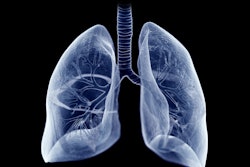
3D reconstruction of CT images could aid in follow-up strategies for pulmonary nodule screening, a study published November 15 in the Journal of Cardiothoracic Surgery found.
Researchers led by Wenfei Xue from Hebei Province General Hospital in Shijiazhuang, China, found through such reconstruction that the tumor blood vessel diameter of ground-glass opacity (GGO) lesions are tied to the growth of malignant pulmonary nodules.
“The identification of ground-glass nodule (GGN) growth is a complicated task,” Xue and co-authors wrote. “However, the pulmonary nodules growth with GGO can be predicted by considering the clinical responses and morphological features in CT images, such as parents’ age and the diameter of tumor blood vessels.”
While blood perfusion has a significant role in assessing tumor progression, this relationship is unclear in patients with persistent pulmonary nodules with GGO. In CT imaging, GGO refers to the increased density inside the lungs. This presents as a foggy or frosty shadow, but not enough shadow to mask the vascular and bronchial texture, the researchers noted.
Xue and colleagues investigated the relationship between tumor blood vessel and the growth of persistent malignant pulmonary nodules with GGO. To reveal the morphological features from collected CT images, the team performed multiplane reconstruction and 3D volume-rendered using PACS system (Neusoft Medical Systems) and a commercially available 3D reconstruction system (InferRead CT Lung, InferVision).
For the study, the team included 116 cases with persistent malignant pulmonary nodules collected between 2017 and 2021. This included 62 patients as stable and 54 patients in the nodule growth group. The team also used multivariate variables logistic regression analysis and Kaplan–Meier analysis for the study.
The researchers found that tumor blood vessel diameter was a significant risk factor in the growth of nodules (p = 0.013). They also found that the cut-off value of tumor blood vessel diameter was 0.9 mm, which included a specificity of 82.3% and a sensitivity of 66.7%.
In subgroup analysis, the team analyzed the ratio between the maximum diameter of consolidation in tumor and the maximum diameter of the whole tumor including GGO, also known as CTR. Through this, the study authors found that tumor blood vessel diameter was significantly tied to the growing processes of nodules (p = 0.027).
The study authors suggested that based on their results, the appearance of the vacuole signs is a direct indicator of tumor growth. This makes continuous monitoring beneficial for timely and effective intervention, they wrote.
“The obtained results are significant for the further clinical decision-making on the management of pulmonary nodules induced by GGN,” they added.
The full study can be found here.


