Radiology researchers are crediting the measurement of cerebral blood flow with laser speckle-contrast imaging -- an invasive alternative to blood-flow imaging with PET or MRI -- for new insights into the triggering of migraine headaches.
Researchers from Massachusetts General Hospital’s NMR Center and Stroke and Neurovascular Regulation Laboratory used their novel imaging approach on rats to confirm brain activity linking migraines to the "aura," or flashing lights, that precede these headaches in many patients.
Specifically, the researchers found that cortical spreading depression (CSD) or electrical depolarization moving across the cortex "activates trigeminovascular afferents and evokes a series of cortical meningeal and brainstem events consistent with the development of headache" (Nature Medicine, February 1, 2002, Vol. 8:2, 136-142).
The findings represent new insight into the pathogenesis of migraines, and new publicity for laser speckle-contrast imaging, an application pioneered by the Massachusetts researchers. Their work will take on clinical significance in the next few months, when surgeons begin using laser speckle-contrast imaging for real-time blood-flow data during brain tumor resection.
Laser speckle-contrast imaging is an open-brain procedure that involves illuminating the cortex with laser light and imaging the resulting speckle pattern. As a result, relative cerebral blood-flow images, with tens of microns spatial resolution and millisecond temporal resolution, are obtained. The regional cerebral blood-flow changes measured with the speckle technique are validated through direct comparison with conventional laser-Doppler measurements (Journal of Cerebral Blood Flow & Metabolism, March 2001, Vol.21:3, 195-201).
Neurosurgeons today generally lack real-time mapping of cerebral blood flow during such operations, said Andrew Dunn, Ph.D., co-author of the Nature Medicine article. The addition of laser speckle-contrast will give surgeons more information for minimizing brain damage as a tumor is removed.
 |
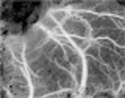
| Speckle-contrast image of cerebral blood flow in rat cortex. Detailed spatial heterogeneities in blood flow can be imaged using this technique. Image courtesy of Andrew Dunn, Ph.D. |
The speckle technique shares its physics with the already available laser-Doppler flowmetry, but provides spatial information the latter does not. Laser speckle-contrast also provides different data than PET or MRI, said Dunn, an assistant physicist at the NMR Center and instructor in the Harvard Medical School department of radiology.
"It has a higher spatial resolution (10 to 25 µm versus 1 mm) and higher temporal resolution," Dunn said, referring to the fact that PET or MRI data would be available minutes after acquisition rather than in real-time.
"The downside of laser speckle-contrast, of course, is that it’s invasive," Dunn noted. "In humans, for now anyway, you would be limited to intraoperative use, so I don’t really see these imaging techniques as competing."
In fact, he added, laser speckle-contrast is complementary to functional MRI. "Here we’re looking at individual blood vessels in the dura, which MR cannot resolve," Dunn said. "But we’re limited to the outermost millimeter of the brain, whereas MR looks below the surface."
One advantage for laser-speckle contrast is its relative ease of use and compact equipment, Dunn said. The technique requires a small laser diode and collimation lens, a camera and camera lens, and a computer to control the equipment. That means that speckle-contrast is "orders of magnitude cheaper and more portable" than MRI or PET, Dunn said.
In the latest migraine study, a 780-nm laser diode and 8-bit CCD camera were placed above the rat’s dura-intact craniotomy to capture a 1.75 x 2.5-mm area. Ten raw speckle images were taken at 15 Hz at five-second intervals over one hour, and a speckle-contrast image was computed for each raw image. Each set of 10 was averaged together and a relative blood-flow image was created by converting speckle-contrast values to correlation times values, the authors wrote.
Laser speckle is "a random interference pattern produced by the coherent addition of scattered laser light with slightly different path lengths," according to the article Dunn authored unveiling the approach. "When an area illuminated by laser light is imaged onto a camera, a granular or speckle pattern is produced."
The quantified blurring of speckle motion can be compared with baseline measurements to see increasing blood flow. In migraines, the increasing blood flow in the dura seen after cortical spreading depression is "presumably the headache," Dunn said.
Laser-speckle imaging was first suggested as a method for blood-flow imaging more than 20 years ago. However, the Massachusetts group was the first to apply the technique in the brain. The speckle technique has also been used to image blood flow in the retina and skin.
Other imaging techniques also laid the groundwork for these latest migraine findings: Functional MRI and PET had previously showed evidence of cortical spreading depression in patients experiencing the pre-migraine aura.
By Tracie L. ThompsonAuntMinnie.com contributing writer
March 19, 2002
Related Reading
Migraine characterized by higher amplitude B waves in middle cerebral artery, April 11, 2001
PET imaging shows distinct pathogenicity of migraine, April 7, 2001
Copyright © 2002 AuntMinnie.com