A vacuum dental cast could be the key to performing the ultimate hat trick in brain tumor treatment, providing a way to fuse multimodality images, guide neurosurgery, and target radiation therapy.
Dr. Reto Bale from the Interdisciplinary Stereotactic Interventional Planning Laboratory at the University of Innsbruck in Austria discussed the results of his group’s work with the Vogele-Bale-Hohner (VBH) mouthpiece at the 2001 European Congress of Radiology in Vienna.
"If a patient comes into the clinic with a suspected brain tumor, CT, MRI, PET, and SPECT scans are performed. Sometimes, stereotactic biopsy is necessary and an invasive frame is attached to the skull of the patient," Bale said. "But then you need additional images, and the frame has to remain in the skull until after the biopsy is performed. You can’t separate the imaging from the intervention."
The VBH mouthpiece (Medical Intelligence, Schwabmünchen, Germany) is attached to the patient’s upper jaw and held in place through pressure. In order to achieve immobilization, the mouthpiece is secured to a base plate by hydraulic arms. A dynamic reference frame provides external fiducials (SIP Laboratory, Innsbruck, Austria).
 |
For image acquisition, the patient is immobilized in the VBH head holder. The mouthpiece is removed and stored in the pre-imaging position. The CT dataset is reconstructed in 3-D and transferred to the frameless stereotactic navigation system. This information determines the entrance and target points of the needle (Radiology, February 2000, Vol.214:2, pp.551-595).
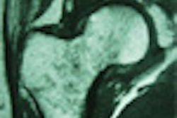
In the first study Bale presented, SPECT and PET images were fused with MRI and CT images in order to combine functional and anatomical information. CT (LightSpeed Plus, GE Medical Systems, Waukesha, WI) and MRI (Magnetom Sonata 1.5 tesla, Siemens Medical Solutions, Iselin, NJ) datasets of 13 patients were superimposed with SPECT images (Vertex V60, ADAC Laboratories, Milpitas, CA) to the desired extent in the axial, coronal, and sagittal planes using a pair-point matching algorithm.
The CT and MR images were fused with a root-mean-square (RMS) error value of 0.8 mm. The CT and SPECT images were fused with an RMS of 2.3 mm. Fusion took anywhere from 5-10 minutes for each data pair. The successful fusing of functional and anatomical images gave the neurosurgeons important information about tumor response and activity, Bale concluded.
 |
For stereotactic biopsy, the VBH mouthpiece was used in a phantom study. A prosthesis of the upper dentition was glued to the underside of the phantom model and its corresponding VBH mouthpiece was created. CT scans with three different slice thicknesses were obtained and registered with the mouthpiece and frame.
The phantom was repositioned and the needle inserted, producing an impression on aluminum foil. Each of the dozen fiducials on the frame was targeted 30 times with 4 steps per scan, totaling 270 punctures. A 3-D localization error vector was calculated for every attempt.
The mean localization error (MLE), which measures the system's degree of accuracy in hitting the target, was as follows:
- 1.84 MLE, 5 mm axial, 0.96 mm standard deviation (SD)
- 1.38 MLE, 3 mm axial, 0.65 mm SD
- 1.31 MLE, 1 mm axial, 0.67 mm SD
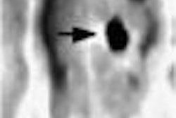
Bale also tested the VBH mouthpiece on 3 cadavers and 7 live patients, all with trigeminal neuralgia. In both the patients and the cadavers, the foramen ovale (3 x 5 mm in diameter) could be reached on the first attempt.
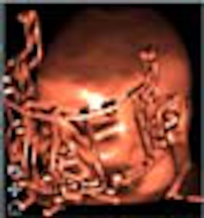
Bale wrote about the group's experience with stereotactic interstitial brachytherapy in Radiology last year. In 12 patients with inoperable cancers of the head and neck, the VBH mouthpiece had a repositioning accuracy of less than 1 mm.
"For intracranial tumors, invasive stereotactic frames screwed into the calvaria have been used for accurate insertion of iodine seeds," he wrote. "In contrast, the VBH head holder allows noninvasive, reproducible, and precise immobilization of the head, which permits temporal and spatial separation of image acquisition, planning, and execution of therapy."
In summary, Bale pointed out the various advantages of using the noninvasive VBH mouthpiece: a single reference system and immobilization device; the ability to fuse anatomical and functional images; accurate targeting for brachytherapy and biopsy; and more accurate follow-up, since the mouthpiece is kept by the patient and can be reused. The next step in the VBH studies will be to determine if the technique is more cost effective than other immobilization methods, he added.
By Shalmali PalAuntMinnie.com staff writer
May 14, 2001
Click here to post your comments about this story. Please include the headline of the article in your message.
Copyright © 2001 AuntMinnie.com