According to neuroscientist and surgeon Dr. Antonio Damasio, the key to consciousness is the awareness of self as different from the world. To him, consciousness is a feeling that accompanies the formation of mental pictures of the individual interacting with the outside world. It is a re-representation of sensory perceptions from the world layered with representations of self as a separate entity. The layers relate the story of what's happening outside to what's happening to the self -- in a kind of homeostatic mechanism that evolved to preserve life by altering the individual's role in it. While the lack of a testable hypothesis has prevented meaningful research in the past, Damasio notes, homeostasis has proved to be a valid basis for investigating the theory with PET imaging.
His 1999 book, The Feeling of What Happens: Body and Emotion in the Making of Consciousness (ISBN 0-15-100369-6), explores the nature of consciousness as it relates to activity in brain regions.
Testing homeostasis
The October issue of Nature Neuroscience (Vol. 3, No. 10) reports on experiments by Damasio and colleagues aimed at investigating the neural basis of emotion and feelings. The team, from the neurology department and PET imaging center of the University of Iowa College of Medicine in Iowa City, studied the brains of 39 normal subjects in experiments aimed at evaluating their recalled emotions with PET.
As hypothesized, researchers found that feeling emotions does require the participation of brain regions such as the somatosensory cortices and upper brainstem nuclei that are known to be involved in mapping and regulating internal organism states. The findings lend credence to the idea that emotions are partly grounded in dynamic neural maps, and thus in homeostasis.
The hypothesis posits that emotions are part of a neural mechanism aimed at maintaining homeostatis, an evolutionary neural mechanism that regulates an organism's current state by executing specific actions through the musculoskeletal system.
In the experiments, four primary emotions -- fear, anger, happiness, and sadness -- were induced by subjects' recall of intensely emotional episodes. The results were then compared with neutral memories of the start of an ordinary day, according to the authors. Subtracting brain activity in a neutral state from those in the emotional state (using a 6 mm Gaussian filter) was assumed to represent the activity caused by emotion. All subjects attained the target emotion during the PET session, with skin-conductance response and heart rate measurements used as a control, they wrote.
PET imaging was done with a GE 4096B camera yielding 15 slices covering a 10 cm axial field of view. Each subject received six injections of 50 m Ci[15O]H2O, two for each target emotion and two for the neutral condition. When subjects signaled with the hand that they had begun to feel the emotion, the radiotracer was injected within five seconds. Dynamic image acquisition enabled reconstruction of the images based on the data collected for 40 seconds after clearance of the arterial blood pool, the authors wrote.
This is your brain on emotion
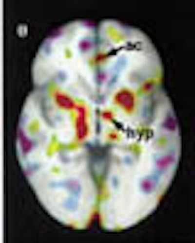
According to the results, activation and deactivation patterns varied qualitatively among emotions. Happiness increased activation in the right posterior cingulate and right SII, while sadness decreased activation in the same regions. The dorsal pons was active in sadness and anger, but not in happiness or fear. The hypothalmus was activated by happiness, anger, and sadness, but showed a negative peak in fear. The insula was active bilaterally in anger and sadness, but only on the right in sadness and fear.
Sadness and happiness caused anterior and posterior activation in the cingulate cortex, yet anger activated only the anterior region, and fear caused negative peaks in the posterior region. The anterior pons showed significant peaks for both sadness and anger, the authors wrote.
The most striking visual results were produced by extreme negative peaks in neocortical areas of both hemispheres, according to the researchers. The decreases were prominent in the left frontal pole for happiness and sadness, and in the right frontal pole for sadness, anger, and fear. Significant decreases were also noted in the right dorsolateral prefrontal regions and the inferior parietal lobule for the various emotions.
The following captioned images illustrate the results:
Figure 1 (197 KB)
Figure 2 (166 KB)
Figure 3 (148 KB)
Other clues
There were a few unexpected results. For example, the amygdala were not highly activated in the tests, a finding that runs counter to previous studies showing the walnut-sized structures to be involved in fear and anger. While the results appear to violate the hypothesis, the authors offered two possible explanations. First, emotions were assessed during the feeling phase rather than the induction phase of emotion in which amygdala are known to be involved. Second, the structures may be less involved in recalled emotions than in actual ones.
The Iowa team also analyzed data from 32 subjects (16 men and 16 women) to assess gender differences, if any, in emotional response. The only significant difference was in the left anterior insula, which was engaged more prominently in women than in men, they wrote.
Interestingly, the patterns for happiness and sadness did not appear to be mere variations of each other, but were qualitatively different in the regions where significant increases and decreases in activity were found, notably the cingulate and orbitofrontal cortex.
Finally, engagement of the pontine nuclei, which receive projections from the cerebral cortex, suggest a whole-body musculoskeletal involvement in the various emotions, the authors wrote.
Overall the results support the hypothesis, they concluded. Emotions do engage structures related to the regulation of homeostasis. At the least, the results indicate a strong connection between emotion and homeostasis, and the activity patterns observed form an important aspect of the mental states known as feelings, they reported.
"We believe these varied patterns provide distinctive perceptual landscapes of the organism's internal state, and that the differences among those landscapes constitute the critical reason why each emotion feels different," Damasio's team wrote. The affected brain regions "work in concert to promote homeostasis, and some do so directly."
By Eric BarnesAuntMinnie.com staff writer
September 20, 2000
Let AuntMinnie.com know what you think about this story.
Copyright © 2000 AuntMinnie.com