A transrectal imaging probe being developed by researchers at the University of Michigan promises both high sensitivity and resolution for early detection of prostate cancer, according to a presentation at the Society of Nuclear Medicine meeting in June.
Lisha Zhang, a research assistant at the university’s biomedical engineering laboratory, laid out the methods and merits of the probe, which is based on the Compton-scatter camera. The Compton-scatter camera is a coded-aperture method that has been used in gamma-ray telescopes, explained Neal Clinthorne, senior associate research scientist in internal medicine, and one of Zhang’s co-authors.
Outfitted with two detectors, the first scatters incident photons while the second detects the scattered photons in time-coincidence. From the energy deposited in each detector, and the positions of the interactions, the incident photon direction can be determined.
In addition, Compton cameras do not have the same restrictions as conventional mechanical or absorbing collimation, which has an undesirable trade-off between counting efficiency and spatial resolution, he said.
The Monte Carlo simulations of the probe consisted of a 1 x 4 x 1 cm3 Si detector with 1 mm3 detector elements. For the purpose of experimentation, the probe was placed at the center of a simulated human body made of a 40 x 40 x 20 cm3 slab phantom with the prostate located 1 cm from the center plane.
Two cadmium zinc telluride (CZT) cameras with the same dimensions as the simulated body were placed 5 cm below and above the body and served as the second detectors.
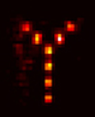
A Y-shaped array of point sources with 5 mm spacing was placed at the prostate location.
The above picture shows the best reconstruction of the simulated prostate at 364 kev. The best reconstruction of the simulated prostate was obtained at 364 kev, which gave 2 mm full width at half maximum (FWHM) resolution at a detection efficiency, including attenuation, of 1.2 x 10⁻³, Zhang said.
At a higher energy of 511 kev, the depth-of-interaction uncertainty in the second detector’s resolution was a limiting factor, Clinthorne said.
While the group is still working on specific tumor detection rates, this initial test shows that the Compton probe offers a high resolution and a high efficiency rate at energies ranging from 140 to 511 kev, which includes the emissions from ProstaScint, a monoclonal antibody labeled with indium-111.
One aspect of imaging with the probe that still requires fine-tuning is the effect of non-prostate background activity on the scan, Zhang said. High energy activity from the spleen, kidney, and liver can interfere with the images.
The group is currently developing a practical Compton-scatter camera for the medical community along with two international companies, CERN of Geneva, Switzerland and IDE AS of Oslo, Norway. A grant proposal to create an intrarectal and external prostate imaging probe, which will be used for patient imaging, has been submitted to the National Institutes of Health, Clinthorne said.
By Shalmali Pal
AuntMinnie.com staff writer
July 6, 2000
Related Reading
Auto co-registration of MR, SPECT images could avoid prostatectomy, June 6, 2000