OSAKA - Breast CAD schemes that hunt for masses and clustered microcalcifications have improved substantially in recent years, and their widespread adoption in the clinical setting is a testament to their growing popularity.
But some researchers think CAD could be a lot better, and potentially find cancers sooner, if more breast CAD systems also looked for architectural distortion -- irregularities in the parenchymal pattern on mammograms without the presence of a visible central mass.
In presentations on Thursday at the Computer Assisted Radiology and Surgery (CARS) meeting, researchers discussed their prototype CAD systems for detecting and evaluating such distortions on mammograms. Their success was dimmed by a relatively high rate of false positives, showing that more work remains to be done. But the groups also found some cancers that mass-based systems overlooked.
First, Takeshi Hara, Ph.D., from Nagoya Bunri University in Japan discussed his group's results with an improved automated detection method based on a pattern distribution assessment in mammography.
The distorted areas on clinical images tend to be of two types, spiculated or retracted, Hara said. While the contour of normal mammary tissue tends to be smooth, the distorted areas are depressed, as depicted in extracted linear data. In the group's previous study, a top-hat processing method for extracting the linear data yielded high sensitivity but too many false positives. The current method attempts to solve this problem by adding dynamic range compression to avoid the effect of steep density changes. The dynamic range compression compresses the pixel value nonlinearly, Hara said.
"The purpose of this work is to develop an automated detection method for architectural distortion on mammograms," Hara said. "To evaluate the detection performance we used a private database and an open database (Digital Database for Screening Mammography [DDSM]) of screening mammograms from the University of South Florida in the U.S."
In a first step, film-screen mammograms are digitized and subsampled to an effective pixel size of 0.2 mm for the detection of architectural distortions, he said. Then the shape of the mammary gland is extracted for shape index and curvedness in terms of shape classes: cup, rut, saddle ridge, and cap. Next, the concentration index is calculated to extract the suspect area. A nine-feature discriminant analysis is performed to eliminate false positives, and the remaining candidates are marked.
The study examined the extraction accuracy in 67 images in terms of shape index and curvedness, mean curvature, and dynamic range compression.
To highlight two points from the complex results, Hara said the training database yielded true-positive results in more than 80% of the cases, with more than two false positives per case. In the testing database (DDSM), true positives stood at 75% with more than 1.5 false-positives per case, he said. The training database contained easier cases.
"The spiculations that were detected only by this method were located around the breast border," the group noted in an accompanying abstract. "It became possible to detect them because the mammary glands around the breast border were sufficiently extracted" by removing the background density through the dynamic range compression step.
The latest approach offered both a higher detection rate and reduced false positives compared to the group's earlier attempts, Hara said, adding that false positives must be reduced further for the system to be clinically viable.
Still, "this detection method could be combined with mass detection without a huge increase in false positives," he said.
Phase portrait method
In a second study presented at the meeting, Rangaraj Rangayyan, Ph.D., and Fabio Ayres from the Schulich School of Engineering in Calgary, Alberta, Canada, developed a slightly different technique to detect architectural distortions, based on the analysis of oriented texture through a shape-constrained phase portrait model.
The group's algorithm uses "a combination of double filters, phase portrait analysis, and several constraints for the reduction of false positives," said Rangayyan, who presented the study based on Ayres' doctoral study.
The most common form of architectural distortion is represented by spiculations arising from a point, Rangayyan said. The second most common form is a focal retraction at the edge of the parenchyma, where tissue strands are pulled, forming a gull wing pattern. In the third type, the incipient mass, tissue components are pushed out.
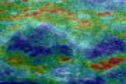
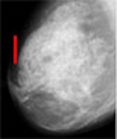
 |
| Architectural distortion in breast tissue (seen in mammographic breast image at right) is a potential indication of malignancy that is often missed by radiologists and CAD systems alike. The distortions can be difficult to appreciate even in the enlarged view (bottom). Images courtesy of Rangaraj Rangayyan, Ph.D. |
 |
"The approach that we have been working on for about four years now is to extract an orientation field, assuming that the breast is made up of oriented texture converging toward the nipple," Rangayyan said.
Their method extracts the orientation field, filters and down-samples it, then analyzes the orientation field using phase portraits. Finally, the phase portrait maps are postprocessed and used to detect sites of architectural distortion.
Mammograms demonstrate oriented textures due to the presence of normal curvilinear structures such as fibrous tissues, vessels, and ducts. In the CAD scheme, the orientation of these structures is obtained by using a bank of Gabor filters as line detectors. Then lines of interest such as spicules and fibrous tissues are separated from confounding lines such as pectoral muscle and parenchymal tissue edges using the orientation field, grading field, and nonmaximal suppression technique.
Having caused a lot of false positives in the past, the confounding curvilinear structures are now removed from further consideration, leaving selected core pixels that can be used in the phase portrait modeling, Rangayyan said.
"The phase portrait of a system of differential equations is the graphical representation of the possible trajectories, in the phase plane, of the state of the related dynamic system," the group explained in an abstract. The phase portrait patterns can be either node, saddle, or spiral as a function of their eigenvalues in a matrix.
"Our task here is to associate the trajectory of the particle under the influence of these velocity components ... and we do that by mapping the oriented field to the orientation patterns," Rangayyan said.
"To analyze a full mammogram, we have a small analysis window, and we slide it over the image," he said. "This is done for every pixel position. Based on the nature of the matrix A, we decide if the pattern is close to a node or a saddle or a spiral, then find the fixed point that may or may not be within the analysis window."
In a small group of 19 cases of architectural distortion from the database (with 41 control mammograms), the group achieved a sensitivity of 84% for detecting architectural distortion with 4.5 false positives per image, "which is fairly comparable to the false-positive rate for the detection of masses in several published studies," Rangayyan said.
The results stand head and shoulders above the group's earlier studies. Before removing the confounding curvilinear structures and implementing other enhancements, previous attempts resulted in as many as 20 false positives per case, he said.
Still, false positives need to be reduced further and the method tested on a larger database, Rangayyan said. The team plans to retrospectively examine mammograms acquired before breast cancer was ultimately diagnosed in screening patients -- to see if the method could have potentially found the cancers earlier.
From the audience, Dr. Robert Schmidt, professor of radiology at the University of Chicago in Illinois, commented on the various architectural distortion schemes presented and the difficulty of acting on the results with or without the aid of a traditional CAD system.
"Clinically this is difficult because we know that architectural distortion for the radiologist is hard to react to anyway -- we miss it," Schmidt said. With CAD, "we have a great deal of difficulty deciding whether to be ... overly reactive, or blind faith or too critical ... and that diminishes the ways we can actually use this. So even if you guys came up with a very good scheme with only a few false positives, we're still going to suffer the problem of how to translate that, so I think we have work to do on this yet."
By Eric Barnes
AuntMinnie.com Staff writer
June 30, 2006
Related Reading
Breast CAD put to the test in high-volume community practice, June 26, 2006
CAD finds breast cancers that radiologists missed for years, March 6, 2006
CAD may cut down on missed breast cancers, January 30, 2006
Breast MRI CAD yields specificity gains, December 12, 2005
CAD falls short of double reading for breast cancer detection, October 25, 2005
Copyright © 2006 AuntMinnie.com