Ultrasound is helping to increase understanding of urinary incontinence and diseases such as urinary bladder carcinoma, both conditions that stand to benefit immensely from improved imaging techniques. Two European studies highlight ultrasound's expanding role in these conditions.
Probing a mystery
Urinary incontinence affects an estimated 200 million adults worldwide, and is considered a public health problem because of its high prevalence among older adults (Nursing Research, November/December 2004, Vol. 53:6, pp. 61-67).
New innovations in ultrasound, though, are helping to demystify the role of the rhabdosphincter in female patients suffering from urinary stress incontinence. Sphincter dysfunction is commonly associated with urinary incontinence.
A multidisciplinary group from the departments of radiology and urology at the Innsbruck Medical University in Austria used an endoluminal color Doppler ultrasound probe (AcuNav, Siemens Medical Solutions, Erlangen, Germany), which enables scanning in the longitudinal plane with a 90-degree sector image, to assess urethral function in 30 women. The study group consisted of healthy women, women with clinically and urodynamically proven stress urinary incontinence, and women who had undergone the sling procedure, a surgical procedure to treat incontinence.
"The currently available probes allow axial imaging only," Dr. Ferdinand Frauscher said in a presentation at the 2004 RSNA meeting in Chicago. Endoluminal ultrasound allows for real-time visualization of the urethral sphincter mechanism, he added.
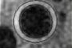
For the intraurethral US exam, the bladder was filled with 250 mL of saline, the catheter was inserted into the bladder, and the external sphincter of the urethra in the area between the middle and distal third of the urethra was identified by its hypoechoic appearance.
The researchers noted there was both constrictive and lengthwise movement of the urethra in the healthy women. "During contraction there is a sliding movement from distal toward the bladder neck," they wrote in their RSNA abstract.
This movement was greatly reduced in the incontinent and postsurgery patients. The mean movement recorded in healthy subjects was 1.1 cm compared to a mean movement of 0.5 cm in incontinent patients, and 0.2 cm in postsurgery patients.
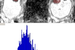
The researchers also used color Doppler ultrasound to assess the resistive index (RI) in the distal, middle, and proximal urethra. The RI findings were compared for the three subject groups.
For all subgroups, the RI decreased from the bladder neck onward toward the distal urethra. For the incontinent subgroup, the RI was lower than in the healthy and postsurgery subgroups.
However, the researchers did not draw any specific conclusions for incontinence based on these findings. "Further studies have to be performed for better understanding of these findings," they stated. But they added that the study results help in better understanding the urethral sphincter mechanism, which in turn is expected to help in developing more and better therapies for patients.
One of the limitations of the study was the small number of subjects -- each subgroup consisted only of 10 subjects. Another limitation was the lack of data about age dependency and factors that may have influenced urethral blood flow. For instance, prior studies have shown that age impacts rhabdosphincter function (Journal of Ultrasound in Medicine, May 2004, Vol. 23:5, pp. 631-637).
In the current study, the subjects in the healthy subgroup (mean age 24.6 years) were younger than the subjects in the incontinent subgroup (mean age 64 years) and postsurgery subgroup (mean age 60.3 years).
3D VEUS
MRI and CT offer high accuracy for staging urinary bladder cancers, especially for invasive cancer. But for local cancers, 3D virtual endocavity ultrasound (3D VEUS) may be the better choice, according to researchers from Russia. They studied the usefulness of 3D VEUS in preoperative staging of bladder tumors.
In men, cancer of the urinary bladder is the second most common cancer after prostate cancer. In women, it is the most common tumor of the urinary tract, according to Dr. Veronika Gazhonova, who presented her group's work at the 2004 RSNA meeting in Chicago.
Gazhonova is the chair of radiology at the Postgraduate Education and Research Center at the President Medical Center in Moscow. She is also the chair of urology with operative nephrology at the Russian State Medical University, also in Moscow.
The patient population consisted of six women and 10 men (mean age 53) with bladder tumors. Transitional cell carcinoma was confirmed in each case through transurethral biopsy. Eleven of the patients had solitary lesions and five had multiple lesions.
The research team used the Aplio system (Toshiba Medical Systems, Tokyo) with high-frequency probes. The probes allowed for differentiation between submucosal, mucosal, and muscle invasion, but was not useful for differentiating deep or superficial muscle infiltration, the authors explained.
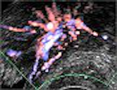
Freehand scanning was used for 3D data acquisition, and surface rendering and multiplanar reconstruction for 3D data analysis. Power Doppler ultrasound was used to differentiate the extent of infiltration. The criteria for staging were based on the vascularity of the tumor's basement. The gold standard was the histomorphology results.
 |
| Criteria for staging on high-frequency ultrasound. T1 = invasion of the mucosa (1) and submucosa (2); presence of hyperechoic rim of submucosa. T2a = infiltration of the muscle (superficial -3); no hyperechoic rim of submucosa; muscle layer is not thickened. T2b = infiltration of the muscle (deep -3); no hyperechoic rim of submucosa; muscle layer is not thickened. T3a = infiltration of the perivesical fat (micro -4); thickened. T3b = infiltration of the perivesical fat (macro -4); thickened muscle layer, irregular external wall. T4 = invasion in the adjacent organs. |
The results from 3D VEUS were compared with pathomorphological results after surgery. The overall sensitivity of 3D VEUS in staging urinary bladder tumors was found to be as high as 91% in the 16-patient study.
 |
 |
| Power Doppler ultrasound criteria for staging vascularity of the tumor basement. Above, submucosal vessels; second, half muscle wall vascular. Below, whole muscle wall vascular; second image, few vessels in the perivesical walls; third image, massive flow in the perivesical fat. Images courtesy of Dr. Veronika Gazhonova, Russian President Medical Center and Russian State Medical University. |
 |
 |
 |
The sensitivity of 3D VEUS for T1 cancers was 75%, and 89% for T2a and T2b cancers. The sensitivity was 100% for T3a, T3b, and T4 cancers. There were three false results: stage overestimation in two bladder neck tumors in T1 and T2a cancers, and stage underestimation in one T2b tumour with distal shadowing.
The findings are significant considering that urinary bladder carcinoma accounts for 70% of malignancies of the urinary tract. Of these, up to 80% are local and only 20% are invasive, Gazhonova said.
The researchers concluded that 3D VEUS was a useful complement to conventional ultrasound, and is sensitive in staging bladder tumors, except in bladder neck tumors and tumors with distal shadowing.
By N. Shivapriya
AuntMinnie.com contributing writer
February 11, 2005
Related Reading
Conservative therapy effective in invasive bladder cancer, December 30, 2004
Adult stem cell injections treat urinary incontinence, November 29, 2004
Copyright © 2005 AuntMinnie.com