B-mode ultrasound has become the gold standard for measuring intima-media thickness (IMT) of the carotid artery. Meanwhile, MRI has proved itself for the noninvasive evaluation of local atherosclerotic disease in the carotid artery.
In combination, these modalities form a powerful duo for assessing plaque morphology and IMT, which is pivotal for successful treatment. Still, one test is always better than two, so a group from the University of Washington in Seattle set out to see if MRI alone could determine carotid artery wall thickness.
Specifically, they used cross-sectional, black-blood, T1-weighted MRI and compared those results to B-mode ultrasound. "If (MRI is) viable, future progression studies would benefit from a reduction in cost, simplification of protocol design, and minimization of patient time," wrote Dr. Hunter Underhill in a poster at the 2005 Society for Cardiovascular Magnetic Resonance (SCMR) meeting in San Francisco in January.
Underhill and co-authors William Kerwin, Ph.D., and Chun Yuan, Ph.D., are from the university's radiology department; co-author Dr. Thomas Hatsukami is from the department of surgery.
 |
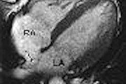
| Example of automated lumen and outer wall boundary detection in a common carotid artery segment (8-12 mm, left to right), proximal to the bifurcation. |
For this study, 17 subjects with 16% to 79% carotid artery stenosis were enrolled. Initial diagnosis was made with duplex ultrasound. The patients then underwent B-mode sonography and MRI, using the technique described above. The MRI acquisition parameters were as follows:
- TR/TE = 800/9.3 msec
- Echo-train length = 8
- Field-of-view = 16 cm
- Matrix size = 256 x 256
- Slice thickness = 2 mm
- Approximate acquisition time = 6 minutes
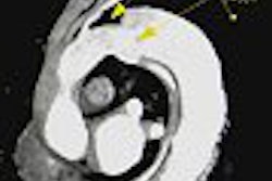
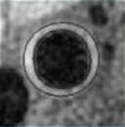
"At a distance 0.8-1 cm proximal to the bifurcation, the lumen and outer wall boundaries of the common carotid artery were automatically detected via a novel technique that utilized a B-spline snake," the group explained. A B-spline snake is a tool for contouring outlining. The mean wall thickness was the average thickness between the two boundaries.
The actual B-spline snake technique that the group used was developed in-house and is currently patent-pending, Underhill wrote in an e-mail to AuntMinnie.com, adding that they hope to make it commercially available in the future.
Ultrasound images were centered 1 cm proximal to the bifurcation. The IMT was measured along the medial common carotid artery wall in the longitudinal plane over a 10-mm length.
The results showed that automated mean wall thickness by MRI was highly correlated with IMT measurements on ultrasound (see graph below). However, MRI did overestimate thickness at lower values. The authors suggested that this may have been because of the adventitia inclusion in the MRI measurement.
 |
| Images courtesy of Dr. Hunter Underhill. |
They concluded that additional testing is required, but also that evaluating mean wall thickness with MRI may serve as an alternative to ultrasound. With MRI, it was possible to obtain local detail and systematic atherosclerotic information in one quick exam, they added.
Underhill clarified a few points to AuntMinnie.com regarding this work-in-progress. First, the six-minute acquisition time is part of a more comprehensive, 30-minute MRI carotid evaluation. In comparison, measuring IMT takes about 15 minutes for both the left and right side.
However, "the advantage of the new MRI alternative to IMT (with ultrasound) is that it can be included in this evaluation with no additional imaging requirements. Essentially, it comes for free for those receiving a carotid MRI," Underhill said. But he stressed that currently, measuring the wall thickness with MRI is only cost-effective if a full carotid MRI scan is planned, thus negating the need for a separate ultrasound test.
"We would have a hard time arguing at this point that MRI should be used when only the IMT is of interest. Unless studies show a better accuracy with MRI, the ultrasound technique will be a faster, lower-cost alternative for measuring only IMT. However, we are currently working toward proving that MRI is more reproducible and accurate, but the data is not finished yet," Underhill stated.
One advantage of MRI over ultrasound is less operator dependence. Underhill pointed out that using MRI for wall thickness relies on a "well-defined and highly reproducible protocol," with similar carotid coverage and image quality across multiple sites and operators. Up-to-date research on reproducibility is slated for publication very soon, Underhill added.
In the meantime, Underhill said the group will continue to work with MRI in this setting. "We believe MRI will prove more reproducible than ultrasound. This will make the added cost of MRI worthwhile. Then, for example, a clinical trial using IMT to gauge results might be accomplished with 100 subjects instead of 1,000."
By Shalmali Pal
AuntMinnie.com staff writer
February 3, 2005
Related Reading
Faster exam time gives CTA a leg up on MRA for peripheral vascular disease, January 19, 2005
Multidetector CT angiography identifies stable carotid plaques, January 12, 2005
Statins do not alter carotid intima-media thickness in diabetics, December 15, 2004
Copyright © 2005 AuntMinnie.com