Down syndrome is the most common chromosomal condition associated with mental retardation, with an incidence in the general population of about 1 in 700. At the 2001 world congress of the International Society for Ultrasound in Obstetrics and Gynecology in Melbourne, Australia, Dr. Beryl Benacerraf discussed her group's research on the use of the genetic sonogram in the perinatal diagnosis of fetal Down syndrome.
Better diagnostic strategies are needed across all maternal age groups. Only 20% of Down syndrome (trisomy 21) fetuses are born to mothers older than 35, and only 20% of Down syndrome children are being identified prenatally, she said.
Benacerraf and colleagues from the Harvard Medical School and Brigham and Women’s Hospital in Boston have developed an ultrasound examination they call a genetic sonogram. The exam, performed between 15-20 weeks' gestational age, identifies specific criteria of markers, which add points toward a sum that indicates the risk for Down syndrome.
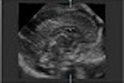
There are six markers in all, including increased nuchal fold, short femur length, short humeral length, echogenic cardiac focus, echogenic bowel, and major abnormality. The nuchal fold measurement, made from the outside of the occipital vault to the skin edge, is abnormally thickened in 40%-50% of fetuses with Down syndrome and carries a very low false-positive rate. Benacerraf said it is the most specific and sensitive marker for Down syndrome.
The team developed a formula for relating the normal fetal biparietal diameter (BPD) to expected femoral length, and found that the optimal cutoff was 0.91 of the expected length. Similarly, a humeral length measurement shorter than 0.9 of the expected length was considered short.
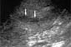
An echogenic focus was present in 18% of Down syndrome fetuses studied, and as an isolated finding it carried a twofold increased risk. Echogenic bowel is a subjective marker, owing to its dependence on transducer frequency and contrast resolution.
Bowel with the same echogenicity as the adjacent bone is considered abnormal. While this abnormality occurs only rarely in the general population (0.5%), it can be seen in 16%-25% of fetuses with Down syndrome, Benacerraf said. In addition, 23% of fetuses with Down syndrome have pyelectasis.
Any heart defect, major anomaly, or increased nuchal fold gives a score of two. One point is added for a short femur, short humerus, pyelectasis, echogenic cardiac focus, and echogenic bowel. A score of one or higher indicates the need for amniocentesis to determine the kayotype, Benacerraf said. When the index score reached two or higher, the method was 73% sensitive for Down syndrome, with a false-positive rate of 4%.
Benacerraf and colleagues analyzed the results of 175 fetuses with Down syndrome, most between 16 and 18 weeks of gestation. Seven hundred control subjects were also included in the study. Looking at nuchal fold measurements of greater than or equal to 6 mm, the sensitivity for Down syndrome was 43%, with a low false-positive rate.
"It is rare to find a thickened nuchal fold in a chromosomally normal fetus," she said.
The probability of disease is highest when the markers appear as isolated findings, and among the markers, thickened nuchal fold is the strongest indicator of disease, she said. A short humerus is the second best indicator, while other single markers are less useful due to their higher prevalence in the general population.
An isolated major anomaly like a heart defect or hydrocephalus has a likelihood ratio of 3, while an echogenic intracardiac focus and pyelectasis carry a likelihood ratio of 1-1.3. The study did not include any Down syndrome fetuses with echogenic bowel as an isolated finding, so Benacerraf said she couldn't comment on likelihood ratios for that finding.
With the right equipment in the right hands, a normal genetic sonogram can mean an 80% reduction of risk for Down syndrome. This equates to a likelihood ratio of 0.2. The likelihood ratios (LR) for the various numbers of markers are -- 0 markers LR 0.2; 1 marker LR 1.6; 2 markers 5.9; and 3 markers 90.6.
"If there (is) more than one marker you can’t start multiplying the individual likelihood ratios; a combination of markers carries a much higher likelihood ratio," Benacerraf said. "So what do you do with a likelihood ratio of one? This means that the established risk is not altered, a 38-year-old remains at the same risk for Down syndrome. You take the patient’s risk from triple screen or even just the age-related risk and this is a most helpful fact -- a marker with a likelihood ratio of one simply doesn’t change the risk."
Benacerraf illustrated how likelihood ratios should be used. If a 39-year-old with a risk for Down syndrome of 1 in 100 has a normal genetic sonogram, the risk for Down syndrome drops to 1 in 333. Her risk is effectively lowered to that of a 34-year-old, and she will probably choose not to have an amniocentesis.
"We should be using the likelihood ratios, rather than alarming our poor patients with any single marker that may occur in a 25-year-old woman who may not really be there at all for the risk of Down syndrome," Benacerraf said.
A 33-year-old with an echogenic intracardiac focus increases her risk from 1 in 400 to 1 in 333, because the likelihood ratio for an echogenic cardiac focus is 1.2. The risk is essentially unchanged with respect to amniocentesis, and Benacerraf questioned whether this information should be shared with the patient, given that the final risk with respect to an amniocentesis remains unchanged.
However, if a 33-year-old has a genetic sonogram showing two markers (LR5.9), her risk rises from 1 in 400 to 1 in 68 -- and the risk is now high enough to consider amniocentesis, she said.
For weighing amniocentesis, Benacerraf said age is no longer an isolated indicator. "Nuchal translucency, genetic sonogram, serum screening in first and second trimester pregnancy, all of these things must be put together so that an individual risk assessment can be derived," she said. The goal is to achieve 90% sensitivity with just a 10% rate of invasive testing.
As in most matters relating to pregnancy, timing is everything. Invasive testing should be performed at 16-18 weeks’ gestation. Nuchal fold measurement must be adjusted for gestational age up to the cutoff of 20 weeks. No genetic sonogram is possible after 22 weeks, Benacerraf said; only a structural survey can be performed after that time.
By Leanne McKnoultyAuntMinnie.com contributing writer
January 4, 2002
Leanne McKnoulty is an accredited medical sonographer currently lecturing in ultrasound at Monash University in Melbourne, Australia. She also is an editor at Ultrasound Review.
Related Reading
Value of genetic sonogram affirmed in reducing need for amniocentesis, June 20, 2001
JUM asserts importance of Down syndrome screening, June 13, 2001
Multinational study makes case for first-trimester sonography, June 12, 2001
Fetal nasal bone examination may improve Down's screening, January 16, 2001
Copyright © 2002 AuntMinnie.com