If researchers at the University of Virginia (UVA) Health System in Charlottesville have anything to say about it, fluoroscopy may one day no longer be needed to guide catheter ablation for atrial fibrillation.
The UVA study team has developed a new catheter ablation technique that combines intracardiac echocardiography and electroanatomic mapping. With an ultrasound catheter, physicians are able to obtain high-resolution images of the heart and other key anatomic structures, according to the researchers.
Complete visualization of the catheter's location is available at all times, allowing physicians to steer the device to the affected target areas. A computer mapping system displays the heart image and the catheter's location in 3D, enabling physicians to record precise location points along the catheter's path, according to UVA.
 |
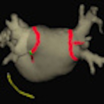
| 3D MRI of the left atrium. The tip of the ablation catheter appears in bright green, and the operator can follow the catheter's movement on the computer screen. This catheter is used to ablate abnormal tissue in the heart. The red dots are the areas that have been treated. |
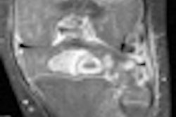
In line with the researchers' goal of eliminating radiation exposure during treatment for atrial fibrillation, cardiac MRI is used for all required imaging prior to the procedure.
In a pilot study of 21 patients with atrial fibrillation, 19 were successfully treated using the technique (Circulation: Arrhythmia and Electrophysiology, December 2009, Vol. 2:6, pp. 611-619).
In the other two cases, two to 16 minutes of fluoroscopy were needed to assist transseptal puncture. The median procedure time was 208 minutes, with a range of 188 to 221 minutes.
 |
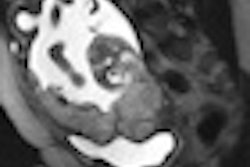
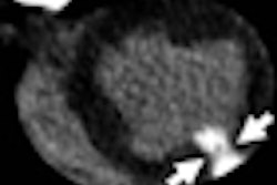
| Ultrasound catheter in the heart. The heart's inner walls appear in white, and blood is black. The catheter's metal tip reflects the ultrasound wave and produces a white circle with a white cone-shaped shadow behind it. The catheter's movement within the heart can be monitored in real-time without the need for x-ray guidance. |
All 21 patients received circumferential pulmonary vein ablation, and eight underwent left atrial ablation. The researchers reported no procedure-related complications.
As a result, the authors found the technique to be feasible and said it merits further research.
"This technique may be especially helpful in preventing x-ray exposure in children, pregnant women, and obese patients undergoing left atrial ablation," wrote the research team led by Dr. John Ferguson.
Larger studies are now needed to confirm the safety of the procedure, according to the researchers. While ongoing investigations are under way, the technique is currently being used in selected patients at the UVA Medical Center to establish the full spectrum of patients who may be able to receive this type of approach in the near future, according to the researchers.
By Erik L. Ridley
AuntMinnie.com staff writer
January 15, 2010
Related Reading
Body mass index predicts left atrial thrombus in atrial fibrillation, December 31, 2009
Copyright © 2010 AuntMinnie.com