Researchers from the University of California, San Diego (UCSD) in La Jolla, CA, are recommending the use of MRI to detect placenta accreta when ultrasound exams prove inconclusive.
The study, presented at the 2009 RSNA annual meeting, found that MRI is a useful and accurate adjunct to ultrasound to diagnose the potentially life-threatening condition. The lead researcher was Dr. Reena Malhotra, a radiologist at UCSD.
Placenta accreta is the normal attachment of the placenta with some degree of invasion into the wall of the uterus. "If it is not diagnosed during pregnancy, it may lead to severe complications at the time of delivery," said co-author Dr. Michele Brown, associate professor of radiology at UCSD. The primary complication includes "massive blood loss by the mother requiring multiple units of transfused blood products."
Placenta accreta is the leading cause of death in women prior to or after giving birth in the U.S. and in developing countries, with deaths occurring in 7% of severe cases, according to Brown.
Increasingly common
The condition also is increasingly common. In 1950, one in 30,000 deliveries was complicated by placenta accreta. The current estimate today ranges from one in 2,500 cases to one in 533 cases. There also are more cases of severe uterus invasion in women who have delivered newborns through cesarean sections, making them more likely to develop placenta accreta.
"The reason that this [condition] can go undiagnosed until delivery is because there are no symptoms during pregnancy in most cases," Brown said. "They may have bleeding occasionally. If it is detected during pregnancy, it is typically found on a prenatal ultrasound."
While ultrasound is the primary imaging modality for placenta accreta, prenatal diagnoses can be challenging. Brown cited previous research that shows that ultrasound's detection rate for placenta accreta varies from 33% to 100%.
Patient sample
 |
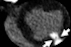
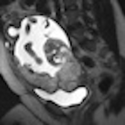
| Above, MR image shows the placenta overlying the cervix with an irregular outer contour and a different appearance of placenta that indicates uterine invasion. Below, image shows the placenta overlying the cervix with a normal, smooth outer contour without evidence of invasion into the uterine wall. Images courtesy of UCSD. |
 |
Surgical and/or pathology results were available in 71 of 108 cases and were retrospectively confirmed with subsequent surgical and pathology findings.
When the researchers correlated MRI results with surgical and pathology findings, MRI demonstrated a sensitivity of 87.8%, specificity of 95.5%, and accuracy of 90.1%. Positive predictive value was 97.7%, while negative predictive value was 77.8%.
The most accurate diagnostic imaging features were abnormal uterine contour and abnormal placental signal, including dark bands on T2-weighted images. For cases in which the MR diagnosis of invasive placenta was uncertain, administration of gadolinium contrast was helpful to distinguish the maternal-placental interface and improve diagnostic confidence.
"Early accurate diagnosis is very important for this condition, because it allows for delivery planning, which improves outcomes," Brown said. Newborn delivery is scheduled and recommended at 36 to 37 weeks, which is early, but complications can increase dramatically after that time.
Based on the study results, the researchers concluded that MRI is a "useful and accurate adjunct to ultrasound for the diagnosis of placenta accreta, which is a potentially life-threatening condition of pregnancy," Brown added.
The researchers also recommended that when there is a high risk of placenta accreta, patients should undergo an ultrasound exam with additional attention given to the placenta. If ultrasound is inconclusive, then an MRI scan should be considered to aid in the diagnosis.
By Wayne Forrest
AuntMinnie.com staff writer
January 6, 2010
Related Reading
Handheld ultrasound performs well in ob/gyn applications, March 14, 2008
US, MR spot life-threatening abdominal pregnancy before it's too late, September 7, 2006
Copyright © 2010 AuntMinnie.com