Researchers at Children's Hospital at Vanderbilt in Nashville, TN, are exploring the characteristics of epiphyseal cartilage perfusion abnormalities among children with osteomyelitis and septic arthritis to help determine better diagnoses for the conditions.
In a study presented at last week's annual RSNA meeting, lead author David P. Johnson, a medical student at Vanderbilt, said researchers found epiphyseal cartilage perfusion abnormalities were more common among children with osteomyelitis and septic arthritis than in a control group.
"The significance of these perfusion abnormalities is unclear," Johnson said. "However, better characterization of these abnormalities may lead to a better diagnosis of osteomyelitis and septic arthritis, conditions that can have a devastating impact on the future of joint development in young children."
Study groups
The retrospective study included children 5 years of age and younger who received gadolinium-enhanced MRI scans between February 2003 and December 2008 at Vanderbilt. Follow-up surgery confirmed cases of either osteomyelitis or septic arthritis after the MRI scan. The control group consisted of children who had no MRI evidence of osteomyelitis and/or septic arthritis.
The MR images were read by a pediatric radiologist who was blinded to the surgical outcomes. The images were reviewed for frequency of focal and global epiphyseal cartilage perfusion abnormalities and correlated with epiphyseal cartilage abscesses or cartilage destruction.
The evaluation revealed 28 epiphyses within the study group and 44 epiphyses among the control subjects. Although both groups had epiphyses with perfusion defects, the study group had 19 epiphyseal perfusion defects, compared with 13 defects in the control group.
Defect details
Among the total number of perfusion defects, there were 11 focal defects (68%) in the study group and 13 focal defects (30%) among the control patients. There were eight global defects (29%) in the study group, but none were found in the control population.
Of the 11 epiphyses with focal defects in the study group, epiphyseal abscesses were present in two cases (18%), while nine epiphyses were normal. For the control group, no abnormalities were found on follow-up of 13 patients with focal perfusion defects
Of the eight epiphyses with global perfusion defects in the study group, a surgically confirmed epiphyseal abscess was present in one patient (12.5%) and epiphyseal cartilage destruction without abscess was present in one case (12.5%). Six epiphyses were normal.
In addition, the researchers found four epiphyseal abscesses and four cases of epiphyseal cartilage destruction in the study group and no abscesses or cartilage destruction among the controls.
 |
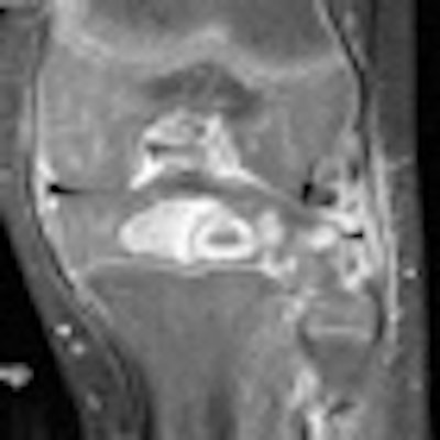
| Image of a 2-year-old with a final clinical diagnosis of proximal tibial epiphyseal osteomyelitis. T1 postgadolinium coronal sequence demonstrates surgically proven intraosseous abscess with diffuse epiphyseal osteitis, surgically proven, with adjacent focal epiphyseal cartilaginous perfusion abnormalities (arrow) and globular areas of abnormal enhancement (arrowhead). Image courtesy of Children's Hospital at Vanderbilt. |
"It is important not to confuse abscesses and perfusion defects despite their similarities, because abscesses must be treated surgically, whereas perfusion defects alone can be followed up over time," Johnson said.
Study limitations
Johnson cited several limitations of the study, noting that control patients with perfusion defects did not have a follow-up surgical exam and not all the patients in the study group with epiphyses received a follow-up x-ray. He added that surgical debridement after MRI, bone destruction, and abscesses likely were confounding factors in findings of growth disturbances at follow-up.
Based on the results, the researchers concluded that perfusion defects are "common in the setting of pediatric musculoskeletal infection, but are also present in controls," Johnson said. "Global perfusion defects are exclusively found on children with osteomyelitis and septic arthritis."
While epiphyseal cartilage perfusion abnormalities are frequent in infection and portend pathology in a minority of patients, the abnormalities for the most part may be related to increased intra-articular pressure and transient epiphyseal ischemia.
And because the perfusion defects may be associated with future growth disturbance, better characterization of the abnormalities could help in diagnosing osteomyelitis and septic arthritis in children, Johnson added.
By Wayne Forrest
AuntMinnie.com staff writer
December 15, 2009
Related Reading
MRI shows prowess in diabetic foot with osteomyelitis, November 10, 2008
FDG-PET may supplement MRI for acute osteomyelitis, August 25, 2008
MRI keeps pace with rapidly evolving musculoskeletal systems of young athletes, May 20, 2007
FDG-PET successfully diagnoses chronic osteomyelitis, July 21, 2006
US overcomes x-ray's limits in pediatric ankle fractures, January 23, 2006
Copyright © 2009 AuntMinnie.com