Orthopedic surgeons are growing more confident in their ability to address labral tears with arthroscopic surgery, but imaging guidance is required beforehand to improve diagnostic accuracy. MR arthrography would be the best bet for assessing these hip abnormalities, according to researchers from Duke University in Durham, NC.
Dr. Glen Toomayan and colleagues recognized that, historically, MRI has been suboptimal for visualizing the acetabular labrum given the latter's small size and variable morphologic appearance. But this shortcoming of MR can be remedied with the right imaging tools, namely a small field-of-view (FOV) and contrast media.
"A good signal-to-noise ratio and excellent spatial resolution are needed for adequate visualization of this small structure," they wrote in the American Journal of Roentgenology. "Labral tears are frequently missed with (MRI) because of a normal, diffusely low-signal-intensity appearance" (AJR, February 2006, Vol. 186:2, pp. 449-453).
The majority of Toomayan's group, including Dr. Nancy Major, is from Duke's radiology department. Co-author Dr. T. Parker Vail is from the division of orthopaedic surgery.
For this retrospective study, 48 patients (51 hips) underwent either conventional MRI (21 hips) or MR arthrography (30 hips) on a 1.5-tesla scanner (Signa, GE Healthcare, Chalfont St. Giles, U.K.). Unenhanced conventional imaging was broken down by a small FOV (14-20 cm) or a large FOV (30-38 cm); all arthrography was performed with a small FOV (14-20 cm).
MR arthrography scanning parameters included spin-echo T1-weighted imaging with fat suppression. The conventional MRI protocol included spin-echo T1-weighted and fast spin-echo T2-weighted imaging.
 |
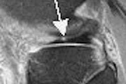
| Axial fat-suppressed fast spin-echo T2-weighted image without intra-articular contrast material (TR/effective TE, 4,000/75; field-of-view, 20 cm) in a 38-year-old woman with hip pain and "locking." Anterior labrum appears diffusely low in signal intensity (arrow) and was therefore interpreted as normal. However, at arthroscopy, patient was found to have anterior labral tear. Toomayan GA, Russell Holman WR, Major NM, Kozlowicz SM, Vail TP, "Sensitivity of MR Arthrography in the Evaluation of Acetabular Labral Tears" (AJR 2006; 186:449-453). |
According to the results, acetabular labral tears were found in 40 of 51 hips at arthroscopy. MR arthrography with a small FOV, and intra-articular contrast, found 24 acetabular labral tears in 30 hips for a sensitivity of 92%. Unenhanced conventional MRI with a large FOV revealed 12 tears in 21 hips for a lackluster sensitivity of 8%. Conventional, no-contrast MRI with a small FOV turned in a sensitivity of 25%.
MR arthrography with a small FOV "allows excellent assessment of the acetabular capsular-labral complex," the group concluded. In addition, "contrast material in the joint can make the distinction more apparent because the contrast material and cartilage are of different signal intensities."
The authors acknowledged that they dealt with a very small patient population, and that additional studies with a larger cohort are needed. However, they reiterated that if the results of future research continue in this positive direction, "an orthopedic surgeon can confidently rely on using MRI to determine which patients will benefit from arthroscopic intervention."
By Shalmali Pal
AuntMinnie.com staff writer
January 24, 2006
Related Reading
X-ray planning improves hip replacement surgical outcomes, January 18, 2006
Study finds MRI is overused in sports medicine, February 24, 2005
MR clarifies paralabral cysts as common source of hip pain, September 6, 2004]
Copyright © 2006 AuntMinnie.com