(Radiology Review) Radiologists at Duke University Medical Center in Durham, NC, recently assessed the prevalence of radial meniscal tears at arthroscopy and whether MR imaging could accurately detect radial tears prior to surgery. Their retrospective study was published in the American Journal of Roentgenology.
Because radial meniscal tear repair is rarely attempted, preoperative diagnosis "can have an impact on operative planning, preoperative counseling, and prognosis," according to Dr. Keith Harper and colleagues. The authors developed four radiologic signs to detect radial tears using MR imaging, including truncated triangle, cleft, marching cleft, and ghost meniscus signs (AJR, December 2005, Vol. 185:6, pp. 1429-1434).
 |
| A schematic diagram showing the marching cleft sign for detecting radial meniscal tears. Harper KW, Helms CA, Lambert HS, Higgins LD, "Radial Meniscal Tears: Significance, Incidence, and MR Appearance" (AJR 2005; 185:1429-1434). |
During a 12-month period, the same surgeon performed knee arthroscopy in 196 consecutive patients and noted any radial tears. Preoperative MR images and reports were reviewed to assess the accuracy of the initial diagnosis. Preoperative MR images were again reviewed using the four specific radiologic signs the radiologists developed for detecting radial tears to assess any improvement in diagnostic accuracy.
 |
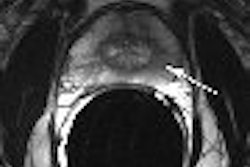
| Marching cleft sign in discoid lateral meniscus indicates radial meniscal tear in 21-year-old man. Conventional sagittal proton density image (TR/TE, 2,000/20) with fat saturation shows partial cleft (arrow) in most peripheral body segment. |
MR imaging was performed using a 1.5-tesla scanner (Signa, GE Healthcare, Chalfont St. Giles, U.K.) with a standard knee protocol. "Sagittal, coronal, and axial fast spin-echo T2-weighted (TR/TE, 4,000/70) and sagittal conventional proton density-weighted (2,000/20) images were obtained" using fat saturation for each sequence, the authors wrote. Two excitations were used for the fast spin-echo images and one excitation was used for the proton density images.
 |
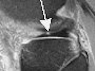
| Marching cleft sign in discoid lateral meniscus indicates radial meniscal tear in 21-year-old man. Conventional sagittal proton density images (2,000/20) with fat saturation show vertical high signal (arrows) extending through adjacent two body segments, indicating cleft marching centrally and anteriorly indicating radial tear. |
Of the 29 patients with radial tears at surgery, 18 had MR imaging available for review. Only seven of 19 tears (37%) shown at arthroscopy were diagnosed on MR imaging preoperatively (one patient had two radial tears). However, using the four signs the authors developed for diagnosing radial tears, they retrospectively identified 17 (89%) of the 19 radial tears found at surgery.
"A more accurate preoperative diagnosis may be rendered using the four described signs to detect radial tears, thus allowing informative preoperative counseling and consideration of new therapies that are available for radial meniscal repair," the group concluded.
The authors noted limitations due to the retrospective nature of their research. However, they were confident that the four signs they described would be more easily understood, recognized, and used by radiologists in the diagnosis of radial meniscal tears.
"Radial Meniscal Tears: Significance, Incidence, and MR Appearance"
Keith W. Harper et al
Department of Radiology, Division of Musculoskeletal Radiology
Duke University Medical Center
Durham, NC 27701, USA
AJR 2005 (December); 185:1429-1434
By Radiology Review
December 23, 2005
Copyright © 2005 AuntMinnie.com