MR spectroscopy (MRS) can keep Alzheimer’s patients and their physicians up to speed as to whether their medication is actually slowing disease progression, according to two reports at the 2003 International Psychogeriatric Association meeting in Chicago.
The goal of these studies was to provide insight into how anti-cholinesterase drugs, such as donepezil, rivastigmine, and galantamine, affect dementia. In the first study, Dr. Maria Caserta and colleagues recruited 31 individuals for proton nuclear MRS scans focusing on three areas of the brain: the right hippocampus, the left hippocampus, and the cingulate gyrus.
Eight of the subjects had been diagnosed with mild cognitive impairment (MCI), while six patients in the study had mild probable Alzheimer's disease. There were 17 control subjects with normal cognition. They were matched for age, sex, and education levels, as the latter is a known risk mediator in Alzheimer's disease.
All scans were performed on a 3-tesla whole-body system (GE Medical Systems, Waukesha, WI) with an appropriate head coil. The scans took approximately 45 minutes.
"We acquired single-voxel proton data in three areas of interest," said Caserta, who is an associate professor in the department of psychiatry at the University of Chicago. "We wanted to use this research tool just to look at the function of the brain, and to specifically use spectroscopy to determine levels of N-acetylaspartate (NAA), a marker of neuronal density."
 |
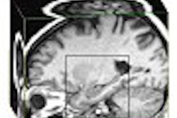
| A 3-tesla proton MR image of a subject with MCI at the level of the mid-hippocampus, with the voxel of interest (2x2x1) placed on the right hippocampus. The left hippocampus was scanned at this level as well. Image courtesy of Dr. Maria Caserta. |
According to the results, the group found distinct and significant differences in levels of NAA. The levels were sharply lower among patients with MCI or probable Alzheimer's disease than with the healthy controls. The effect was especially apparent in the right hippocampus. They found no substantive differences in the posterior cingulate gyrus.
Caserta said the differences in the right and left hippocampal regions were intriguing because they suggested that the beginnings of dementia might originate in the hippocampus even before the appearance of cognitive symptoms.
For example, the NAA/creatine ratio in the right hippocampus was 1.45 for normal controls, 1.12 for those with MCI and 1.18 for patients with early Alzheimer’s disease. In the left hippocampus, the ratio was nearly identical for the controls, 1.46, but declined to 1.33 in those with MCI and 1.21 in the Alzheimer’s disease population.
 |
| A 3-tesla proton NMRS scan obtained from a similar right hippocampal voxel as seen above. The NA:Cr ratio, as well as the Ch:Cr and mI:Cr ratios, are shown on the right-hand side. Hippocampal spectroscopy scans were nine minutes for each hippocampus voxel. The posterior cingulate gyrus scan (data not shown here) was approximately two minutes. Other parameters include a TR=2000 and a TE=30. Image courtesy of Dr. Maria Caserta. |
Commenting on the study, Dr. Stephen Salloway said he had reservations about the study's small patient population, but added that "this data appear promising and might be interesting to use as outcome measures in clinical trials."
Salloway, who is an associate professor of psychiatry at Brown University in Providence, RI, added that one of the tools needed to expand the use of cholinesterase inhibitors is a way to measure, on an individual basis, if the medication is working.
Imaging for outcome measures
In a second study, Dr. K. Ranga Krishnan and colleagues used MRS to measure levels of NAA in the brain structures of patients with Alzheimer's disease being treated with either donepezil or a placebo.
"Alzheimer's disease is associated with atrophy of the temporal lobe structures," said study presenter Dr. P. Murali Doraiswamy, who is chief of the division of biological psychiatry at Duke University in Durham, NC.
 |
| Based on MRS results, donepezil increased brain NAA levels between weeks 6 and 18, and slowed the progression of hippocampal atrophy over the duration of the study. Image courtesy of Dr Cecil Charles, Duke Image Analysis Laboratory. |
This study used MRS, MR volumetrics, and PET to ascertain hippocampal volumetric measurements in 34 patients on donepezil and 33 patients receiving placebo. There was a decrease in volume of 8.23% among the patients getting placebo, versus a 0.37% decline in volume among those receiving donepezil.
"We are now seeing that these different imaging modalities are being used to look at surrogate markers in determining treatment outcomes in Alzheimer's disease," commented Dr. Gary Small, director of the Neuropsychiatric Institute of the UCLA Center on Aging in Los Angeles.
 |
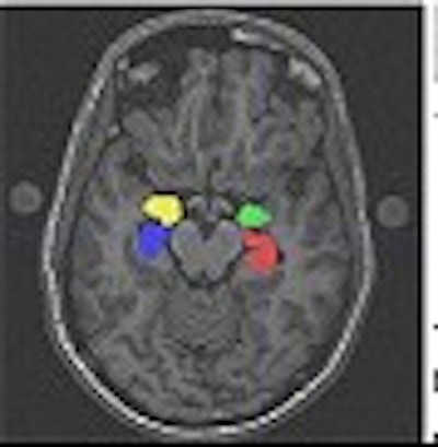
| Three orthogonal sections of a high-resolution T1-weighted MRI image with color overlay of segmented hippocampus and amygdala. The fourth window displays the anatomical objects in three dimensions, and facilitates verification of slice-by-slice processing of 3-D objects. The segmentation and visualization tools are available for free at http://www.cs.unc.edu/~gerig. Image courtesy of Dr. Guido Gerig. |
Small said that technological developments over the past decade have led to a greater understanding of brain function and genetic risk in Alzheimer’s disease, particularly in the early stages.
For assessing dementia or age-related memory complaints, functional imaging offers the advantage of providing a positive diagnosis of early Alzheimer’s disease, often before clinicians can identify the condition using conventional clinical assessments. In addition, evidence is accumulating that nonpharmacological interventions may decelerate brain aging, Small said.
By Edward SusmanAuntMinnie.com contributing writer
August 12, 2003
Related Reading
Loss of brain volume on MR may precede clinical signs of Alzheimer’s, September 5, 2003
Reference database of brain images goes online, August 8, 2003
Scans suggest why education prevents Alzheimer's, August 8, 2003
Alzheimer's brains show features of immaturity, May 8, 2003
Imaging's role still evolving in dementia diagnosis, April 29, 2003
Copyright © 2003 AuntMinnie.com