Dynamic contrast-enhanced MRI can differentiate benign from malignant breast lesions in women with mammographically detected microcalcifications, perhaps sparing them the need for surgical biopsy.
Using both signal intensity and visual interpretation techniques in MRI, researchers significantly bolstered accuracy in the evaluation of suspected ductal carcinoma in situ (DCIS), according to a study from the University of Hull in Kingston-upon-Hull, U.K. The findings were reported last month at the annual meeting of the 2002 American Society of Clinical Oncology in Orlando, FL.
MRI has been considered unsuitable for the assessment of microcalcifications, as the spots of calcium are often tiny enough to escape detection. However, dynamic contrast techniques rely on a different approach, highlighting breast tissue that has increased microvessel density as a result of angiogenesis.
"DCIS has been shown by several groups to induce angiogenesis that can be demonstrated by gadolinium contrast as a focal or branching pattern of contrast uptake, and therefore we do not need the calcium to see the in situ disease," said Dr. Peter Kneeshaw, lead investigator.
The study enrolled 71 women aged 36 to 83 with microcalcifications of the breast. Imaging was done both without contrast and after intravenous infusion of 0.1 mmol/kg gadolinium, in order to track changes in signal intensity over time. Lesions were evaluated morphologically as well.
Using a 1.5-tesla scanner, a bilateral phased-array breast coil, and fast spoiled gradient-echo sequences, researchers acquired 35 images in four slice locations in the sagittal plane in areas characterized by parenchymal deformity on noncontrast imaging. If no deformity was noted, they acquired 35 images in nine slice locations in the coronal plane over the entire breast, all in an area classified by mammography as suspicious or indeterminate.
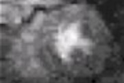
The video clip below is of a dynamic MR examination. It shows one slice location with 35 images shown in sequence, the gadolinium contrast been given after the second set of images. The area is high grade DCIS associated with
microcalcification. Note the rapid uptake of contrast.
![]()
All women had core biopsy or fine-needle aspiration. Five were found to have invasive disease, 14 had DCIS, and 18 had both invasive disease and DCIS. In the remaining 34 women, the lesions were benign.
When pathologic findings were compared along with various characteristics of signal intensity, MRI was found to successfully distinguish malignant from benign lesions. For example, the enhancement index at one minute was 0.48 for malignant lesions and 0.23 for benign lesions.
Similarly, signal amplitude in the most-enhancing nine-pixel square was 7.59 for malignant lesions and 2.08 for benign lesions. Both differences were highly statistically significant. The sensitivities for malignancy of these two signal intensity measurements were 71% and 90%, respectively, and their specificities were 83% and 75%, respectively.
 |
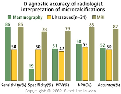
In addition, radiologists were more accurate in their interpretation of dynamic contrast-enhanced MRI when compared to other imaging methods (see table above). For example, mammography rated a sensitivity of 86%, a specificity of 19%, and an overall accuracy of 52%. By comparison, the sensitivity of contrast-enhanced MRI was 86%, the specificity 78%, and the accuracy 82%. Ultrasound fared worse, achieving only 50% in all three measures.
Kneeshaw and his colleagues concluded that dynamic contrast-enhanced MRI is superior to other imaging modalities when used to assess microcalcifications.
"It may offer an alternative to open surgical biopsy for women with equivocal findings following assessment of clinically occult disease," he said.
By Catherine CarringtonAuntMinnie.com contributing writer
July 22, 2002
Video clip courtesy of Dr. Peter Kneeshaw.
Related Reading
MRI successfully tracks women with hereditary breast cancer risk, April 8, 2002
MRI beats US, mammo for tracking breast tumor response, January 7, 2002
MRI proves sensitive for high-grade DCIS in breast cancer, January 2, 2002
Patients with known breast malignancy benefit from MRI, November 25, 2001
Copyright © 2002 AuntMinnie.com