Depending on what part of the world you’re in, rabies can be a significant menace. Globally, the number of human deaths due to rabies is estimated at 35,000 and 50,000 annually, according to the Rabnet data bank maintained by the World Health Organization.
The occurrence of rabies in humans varies wildly: In 2000 in the U.S., five cases were logged with Rabnet. By comparison, in 1998, India reported 31,300 instances of rabies in humans.
Faced with so many cases, doctors in India have found a role for MRI in diagnosing radiculomyelitic rabies. The modality can be useful in differentiating between radiculomyelitic (also known as silent) rabies and disseminated encephalomyelitis, which has similar clinical presentations.
Rabies is a zoonotic, viral disease that infects domestic and wild animals and is transmitted through close contact with saliva from the infected creature. The initial signs of rabies infection can either be hyperactivity (furious rabies) or paralysis (silent rabies). In furious and dumb rabies, coma and death, generally due to respiratory failure, follow complete paralysis.
"Silent rabies is about 25% of the rabies that we see. The number of rabies cases that we see every year is 5-6 in children and 10-12 in adults," wrote Dr. Sunit Singhi in an e-mail to AuntMinnie.com.
Singhi is an additional professor in the department of pediatrics at the Postgraduate Institute of Medical Education and Research (PGIMER) in Chandigarh, India. Singhi and his co-authors from the departments of radiodiagnosis and pathology -- Drs. Ravi Desai, Vivek Jain, Paramjeet Singh, and Bishen Dass Radotra -- wrote about their experience with MRI and rabies in the American Journal of Neuroradiology.
The disease is most commonly transmitted through wild animal bites, including raccoons, skunks, bats, coyotes, foxes and some domesticated animals, such as dogs.
In one case report, a 10-year-old boy presented at PGIMER with a history of fever, progressive weakness of both lower limbs, and extreme agitation with behavioral changes. The patient had been bitten on the left lower limb by a stray dog 25 days earlier; 15 days later, he was given a course of Rabipur vaccine but did not receive anti-rabies immunoglobulin, the group stated (American Journal of Neuroradiology, April 2002, Vol.23:4, pp.632-634).
"An examination performed at admission revealed that the child was in altered sensorium (Glasgow Coma Scale score of E3M4V2). CT of the head was performed at admission, and the results were normal," they said. However, MRI revealed less encouraging results.
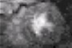
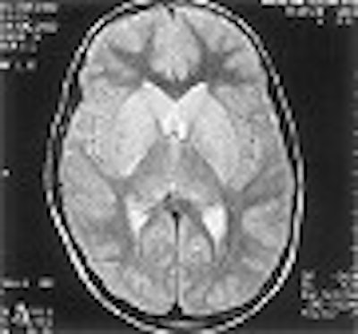
MR imaging was performed on a 1.5-tesla Magnetom Vision Plus scanner (Siemens Medical Solutions, Erlangen, Germany). Axial T2-weighted images showed hyperintensity involving central gray matter of the whole spinal cord, extending cranially to symmetrically involving the bilateral thalami, hippocampi, basal ganglia, and the dorsal aspect of the brainstem. A T2-weighted image at the level of the basal ganglia revealed hyperintensity of the caudate head and lentiform nucleus; another T2-weighted image done at the level of the pons showed hyperintensity of the posterior portion of the pons.
 |
| A T2-weighted MR image of the brain (A), scanned at the level of basal ganglia. The exam reveals hyperintensity of the caudate head and lentiform nucleus. In image B, the T2-weighted MR image obtained at the level of the pons reveals hyperintensity of the posterior portion of the pons. Images courtesy of the Dr. Sunit Singhi. |
Because of the MR results showing the involvement of gray matter, the administration of steroids was discontinued. Life support was withdrawn at the family’s request 10 days after hospital admission.
"Rabies has an affinity for bulbar and limbic neurons and specifically involves the brainstem," the group wrote. "In the cases of silent rabies, the most striking pathologic changes are seen in the spinal cord...MR images showed the predominant gray matter involvement of brain and spinal cord, which is a hallmark of rabies and an important differentiating point from acute disseminated encephalomyelitis."
The hyperintensity of spinal cord gray matter on the T2-weighted images corresponds to inflammatory cell infiltration, they concluded.
MR is now routinely done at PGIMER to confirm diagnoses, and "has helped us in saving the patient with demyelinating encephalomyelitis," Singhi said.
Another group, from King Edward VII Memorial Hospital in Mumbai, India, had similar success with MR for diagnosing rabies encephalitis. In this case, a 30-year-old man presented with a variety of symptoms, including altered consciousness and fever, after being bitten by a dog. Axial spin-echo T1-weighted MR scans showed hyperintensity in the globus pallidus and putamen bilaterally. Axial spin-echo proton density-weighted MR scans showed hyperintensities in the heads of the caudate nuclei bilaterally (AJNR, April 2001, Vol.22:4, pp. 677-680).
"Paralytic rabies encephalitis was diagnosed and subsequently established ante mortem...the patient gradually deteriorated and died within three days of admission," wrote Dr. Manasi Awasthi and colleagues.
They concluded that while the diagnosis of rabies is mainly clinical, patients may present with a number of symptoms, and that in the appropriate context, MRI can narrow that diagnosis down.
By Shalmali PalAuntMinnie.com staff writer
July 22, 2002
Related Reading
MRI trains an eye on orbital Lyme disease, June 10, 2002
Copyright © 2002 AuntMinnie.com