Some thyroid cancer patients with elevated serum thyroglobulin (Tg) levels may not benefit from conventional iodine therapy. However, FDG-PET can detect the tumors 131I imaging misses, according to French researchers.
A study in the latest issue of the Journal of Nuclear Medicine assessed the clinical impact of 18FDG-PET on patients with differentiated thyroid carcinoma who underwent therapy with 131iodine, yet still had elevated Tg levels as well as negative 131I whole-body scans.
"After the ablation of residual thyroid tissue, measurement of serum Tg levels and 131I imaging usually provide sensitive tools for the early detection of recurrence. Unfortunately, a few patients...even after administration of therapeutic doses, [have] an inability to trap iodine," wrote lead author Dr. Badia O. Helal from the Hôpital Antoine Béclère in Clamart. The other authors hailed from various French institutions, including the Hôpital Saint-Louis in Paris, and the Institut Jean-Godinot in Reims (JNM, October 2001, Vol.42:10, pp. 1464-1469).
The study sample consisted of 37 patients who were evaluated with FDG-PET after initial treatment for thyroid carcinoma which included total thyroidectomy and 131I ablation therapy. The pathologic tumor stage for the majority of the patients (18) was T4. A clinical stage III was given to 16 patients.
A few days after the patients received the therapeutic dose of 131I, whole-body scanning was performed using a dual-head, large-field-of-view gamma camera with a high-energy collimator. Additional conventional imaging (CI) included chest x-ray, sonography, and thoracic CT.
"The patients were divided into 2 groups: group 1, which consisted of 10 patients with positive CI results, and group 2, which consisted of 27 patients with negative CI results," they wrote.
The PET scan was performed 50-60 minutes after injection of 370 MBq FDG, and imaging was done on an ECAT Exact HR+ scanner (Siemens-CTI, Knoxville, TN) with an axial field of view of 15.5 cm. All scanning was performed in 3-D mode.
 |
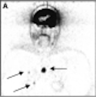
According to the results, FDG-PET findings were positive in 76% of the patients in group 1. The scan confirmed an additional 11 tumor sites, including cervical and mediastinal lymph nodes and distant metastases. In group 2, FDG-PET identified 44 tumor sites affecting a number of areas, such as the neck and mediastinum.
Finally, the scan was negative in 8 patients with persistent elevated Tg values. These cases have been continually followed up and remain without detectable metastases.
"For all types of cancer, the detection rate was significantly higher for stages III and IV (80%) than for stages I and II (47%)…The PET findings led to a change in the management of 29 patients," the authors said.
 |
One of the benefits of PET scanning over other modalities is its ability to perform a whole-body exam with great spatial resolution for higher sensitivity. In addition, the authors found a correlation between FDG uptake and the stage of thyroid cancer in both papillary carcinoma and follicular carcinoma.
"We suggest that FDG-PET be used as a first-line investigation in patients with negative 131I post-therapy scan results and elevated Tg levels," they concluded.
By Shalmali PalAuntMinnie.com staff writer
October 3, 2001
Related Reading
FDG-PET influences thyroid treatments, January 18, 2001
Copyright © 2001 AuntMinnie.com