SAN FRANCISCO - Pulmonary embolism (PE) presents a litany of diagnostic challenges, and radiologists have tackled them with everything from angiography to CT, MRI, and scintigraphy.
The smallest clots are the hardest to find, of course, and despite advances in CT and MRI techniques, neither of these modalities can reliably target emboli in the smaller vessels. Moreover, interpreting the scans is still a variable and subjective process that requires great skill on the part of the radiologist.
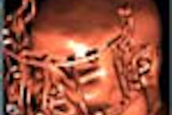
Against this backdrop, researchers from the University of California, San Diego Medical Center have found initial success with a new monoclonal antibody-based radiopharmaceutical that binds to even the tiniest emboli. Dr. Timothy Morris and colleagues used the technetium-tagged radiotracer in conjunction with SPECT to detect PE induced in dogs.
In 35 subjects so far, the agent has never failed to detect PE, and has never accumulated in nonthrombotic regions said Morris, who is associate professor of medicine at UCSD, and director of the acute venous thrombo-embolism service at the center. He discussed his poster presentation with AuntMinnie.com at this week's American Thoracic Society meeting in San Francisco.
"The idea is that we wanted to combine two different techniques to make images of relatively small PE, because that's where the major diagnostic gap is right now: we can't find small emboli," Morris said. "So we used a monoclonal antibody that is raised against part of the fibrin molecule. [The antibody] is only formed when a clot is formed, so it's a constituent that's not found anywhere else in the body besides in blood clots."
Fifteen mCi of Tc-99m is attached to anti-fibrin antibody fragments known as FAB' (Agen, Brisbane, Australia), and injected into the subject. The solution clears the body fairly quickly, yet binds to the clots, which are then detected with SPECT about four hours later, Morris said.
The study in the poster presentation used a human/murine chimeric antibody fragment in three canine subjects, but since then the team has found better results with a slight variation of the compound. FDA approval hasn't been applied for, but the researchers hope to begin clinical trials in about a year, Morris said.
"We've worked with different variations of the same antibody, different chain lengths, and this [chimeric] fragment with [15 mCi] of technetium seemed to work out the best," Morris said. "Since then, we've had much better results with a deimmunized version."
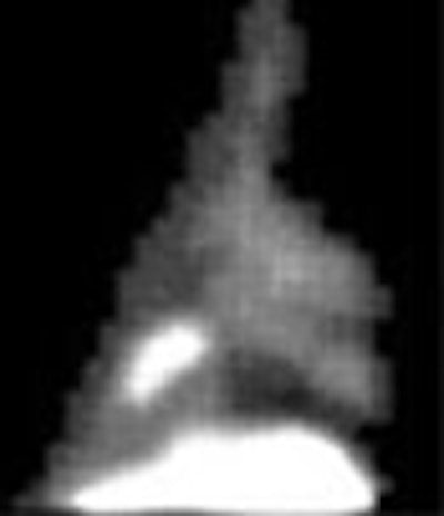
In the poster study, PE was induced in three dogs by infusing thrombin and fibrogin through balloon catheters placed in the femoral veins. One hour after embolization, 15 mCi of Tc-99m-labeled FAB' was injected in the femoral veins of the anesthetized subjects. Three pulmonary emboli were induced in the subjects, with masses of 0.45 g, 1.9 g. and 0.09 g, respectively. All 3 emboli were intensely radiolabeled, with clot/blood radioactivity ratios of 27.2, 38.1, and 45.1, respectively.
SPECT images were acquired 8 hours following injection of the agent (the delay has since been reduced to 4 hours), and reconstructed using standard techniques. Fifteen images of the thorax were created in 24º increments and displayed rotationally for each subject. The animals were subsequently destroyed and autopsied, and the emboli were extracted and weighed.
According to the results, all 3 emboli were visualized as "hot spots" on SPECT. The study concluded that the method produced clear images of pulmonary emboli, even peripheral emboli of relatively small size. The technique is also presumed, though not yet proved, to allow simultaneous imaging of deep venous thrombi (DVT), Morris said.
The technique requires no breath-holding, fasting, or nephrotoxic contrast agents. And because scan results are easy to read, the method could significantly reduce the interreader variability that can occur with other techniques, Morris said.
 |
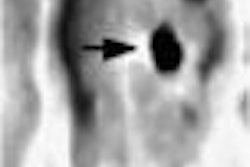
"These emboli are about a tenth of a gram large," Morris said, noting darkened areas on the poster images. "There's no way angiography, helical CT, etc. can find clots this small. This one (shown above) is only a half a gram. This is about as small a clot as would be clinically important, and we can image it beautifully."
Morris said portability was another potential advantage, since the agent could conceivably be injected in the emergency room -- or anywhere -- and the patient taken to nuclear medicine a few hours later for scanning.
"This technique has the promise of making every hospital a center of excellence for PE," Morris said. "There will be no longer be a dependence on one skilled radiologist in every city to be able to read the scan -- everyone should be able to read the scan."
By Eric BarnesAuntMinnie.com staff writer
May 25, 2001
Related Reading
D-dimer assay rules out PE, DVT in emergency triage, March 27, 2001
Negative CT angiography sufficient to rule out pulmonary embolism, March 9, 2001
Serial ultrasound not cost-effective for diagnosis of deep vein thrombosis, March 1, 2001
Spiral computed tomography effectively rules out pulmonary embolism, February 12, 2001
Study claims that ER doctors are too cavalier about ordering V/Q scans, November 28, 2000
Click here to post your comments about this story. Please include the headline of the article in your message
Copyright © 2001 AuntMinnie.com