The response to therapy in solid breast tumors is currently evaluated by measuring the change in tumor size. Unfortunately, this method is often inaccurate during the early stages of chemotherapy. Properly identifying tumor response in the early stages is vital, either to determine if additional courses of therapy are needed or if treatment should be discontinued.
A group of gynecologists and radiologists have proposed using sequential F-18 FDG-PET for the early prediction of chemotherapy response. Dr. Joerg Dose Schwarz and colleagues also hypothesized that changes in the pattern of FDG uptake would correlate with overall survival rates.
"Modern treatment regimens in metastatic breast cancer are developed toward being tailored to the needs of each patient," they wrote. "Early identification of nonresponders is crucial to avoid ineffective treatment, unnecessary side effects, and costs" (Journal of Nuclear Medicine, July 2005, Vol. 46:7, pp.1144-1150).
Schwarz and three of his co-authors are from the department of gynecology at University Hospital Hamburg-Eppendorf in Hamburg, Germany. Co-author Dr. Lars Jenicke is from the hospital's nuclear medicine department. Co-investigator Dr. Norbert Avril is from the University of Pittsburgh Medical Center.
For this study, 11 patients were enrolled. They were already in a phase III chemotherapeutic clinical trial, comparing epirubicin/cyclophosphamide to epirubicin/paclitaxel. Treatment was repeated every three weeks, up to a maximum of 10 cycles. The women first underwent conventional imaging (ultrasound, CT, MRI), which identified a total of 25 separate metastases. FDG-PET scans were repeated after three cycles at nine weeks, six cycles at 18 weeks, and nine cycles at 27 weeks.
Imaging was done on a whole-body PET scanner (ECAT EXACT 47/921, CTI/Siemens Medical Solutions, Erlangen, Germany) after the IV injection of 240-400 MBq of FDG. A nuclear medicine physician who was unaware of patient details interpreted the PET images.
Based on conventional imaging, six out of 11 patients were classified as responders and five as nonresponders after chemotherapy completion. These results were considered the gold standard.
Of the 26 lesions, 17 responded to chemotherapy. On FDG-PET, standard uptake values (SUVs) decreased to 72% after the first cycle and 54% after the second cycle when compared with the baseline PET scan. In nonresponders (nine lesions), the SUVs only declined to 94% after the first cycle and 79% after the second. This difference was statistically significant, the authors stated.
 |
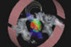
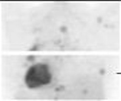
| A patient with partial response to palliative chemotherapy. Image courtesy of Dr. Joerg Dose Schwarz. |
"F-18 FDG-PET correctly predicted the response in all patients as early as after the first cycle of chemotherapy," they wrote. "F-18-FDG-PET was superior specifically in the identification of nonresponders." The modality pinpointed three nonresponders after one cycle of treatment and three nonresponders after four cycles.
The mean follow-up time was 14.5 months during which 10 patients died. The overall survival in nonresponders was 8.8 months versus 19.2 months in responders.
The authors acknowledged that their study was small, that they did not perform a quantitative SUV analysis, and that FDG-PET was not used to change patient management. However, they stated that FDG-PET did have more promise than conventional imaging for assessing these patients. Future studies will have to establish a threshold for differentiating between responders and nonresponders on PET.
By Shalmali Pal
AuntMinnie.com staff writer
August 17, 2005
Related Reading
Ultrasound useful for evaluating early-stage breast cancer after chemo, July 6, 2005
Breast MR accurately predicts tumor response to chemo in two studies, June 14, 2005
PET sharpens treatment plan for advanced breast cancer, August 16, 2004
Copyright © 2005 AuntMinnie.com