NEW ORLEANS - Myocardial uptake of FDG following an ischemic episode may serve as a memory marker of transient ischemia, resulting in a more sensitive and specific way to detect acute ischemia, according to results of a pilot study presented this week at the American College of Cardiology meeting.
"The main implication of the study is that it proves the feasibility of 'hotspot' FDG imaging to ischemia after stress. The importance of the hotspot feature is that, when we're looking at ischemia (with) current technology, we're looking at defects," explained co-author Dr. James Arrighi.
In this study, "rather than looking at defects, we're looking at uptakes in the myocardia as a specific metabolic signal of ischemia, which potentially has the capability of being more sensitive and specific for ischemia detection," added Arrighi, who is an associate professor of medicine and diagnostic radiology at the Yale University School of Medicine in West Haven, CT.
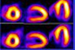
 |
| Image courtesy of Dr. Brian Abbott. |
For this preliminary investigation, ten male patients with mild to moderate ischemia (mean age 69 years; seven with known coronary artery disease), underwent exercise SPECT myocardial perfusion imaging (MPI) with repeat exercise testing within one to two weeks. The patients then had an FDG-PET scan, one hour post-exercise.
"SPECT and PET images were assessed visually and quantitatively using regions of interest for heart:lung ratios and a circumferential profile-based analysis modified for ‘hotspot’ imaging. A change in regional maximal uptake, >10% on SPECT, and regional FDG PET uptake, >110%, was considered as evidence of ischemia," according to the researchers.
The results showed that five out of ten patients had enhanced regional FDG uptake. The mean heart-to-lung ratio was 5.3 in visually abnormal uptake versus 3.0 without visual uptake for a p=0.02.
Overall, they noted that eight out of ten patients had evidence of FDG uptake by visual or quantitative analysis. All patients with a visually or quantitatively positive FDG scan had uptake in either an ischemic SPECT region or a territory with known coronary artery disease by angiography, researchers stated.
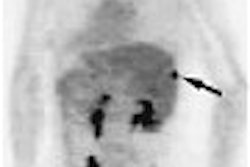
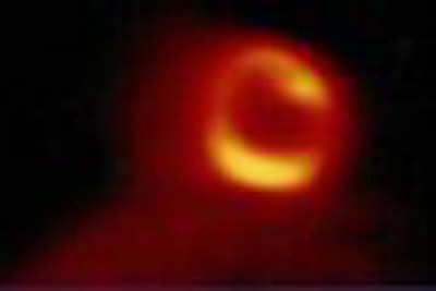
 |
| Image courtesy of Dr. Brian Abbott. |
"We have data from our institution that shows that during an episode of myocardial ischemia the heart can actually switch over from fatty acid metabolism to glucose metabolism. One way to take advantage of that metabolic switch would be to image glucose uptake in the heart as a mark of ischemia, using FDG," said presenter Dr. Brian Abbott with the VA Connecticut Health Care System in West Haven, CT, and an assistant professor of medicine at Yale.
"If FDG could be imaged after a brief ischemic episode, 60 to 90 minutes later (a time point that may be clinically relevant), and if we were to use FDG as a way to image a preceding episode of ischemia -- such as someone presenting in an emergency department with acute chest pain -- it could be used as an alternative to exercise perfusion imaging, in that it would be a hotspot way of imaging ischemia, which could be incorporated into the protocol for stress testing," Abbott said.
Although the results of the research do present new clinical possibilities, FDG-PET is not quite ready to replace exercise SPECT, the authors cautioned. Further research is needed to assess the validity of the study.
By Jerry IngramAuntMinnie.com contributing writer
March 9, 2004
Related Reading
Ultrasound boosts efficacy of TPA for ischemic stroke, February 10, 2004
Myocardial ischemia, viability made apparent with MR, February 6, 2004
PET may overpredict myocardial recovery compared with MRI, March 11, 2003
Copyright © 2004 AuntMinnie.com