Adding FDG-PET imaging to CT for ovarian cancer staging can aid in the detection of metastases outside the pelvis -- an important consideration when deciding whether cryoreductive surgery alone will be effective. A multidisciplinary group from Fukui Medical University in Fukui-ken, Japan studied the incremental benefits of using FDG-PET and CT in patients with tumor spread.
"Despite advances in surgery, complete removal of the tumor is still impossible in approximately 60% of patients with ovarian cancer," wrote Dr. Yoshio Yoshida and colleagues. "The accuracy of CT in staging of ovarian cancer has been reported to range from 60% to 80%" (American Journal of Roentgenology, January 2004, Vol.182:1, pp. 227-233).
Yoshida is from the university’s department of obstetrics and gynecology; his co-investigators were from the departments of neurosurgery, radiology (Dr. Tatsuro Tasuchida), and the Biomedical Imaging Research Center.
For this study, 15 patients underwent abdominopelvic CT and whole-body FDG-PET two weeks before full pelvic and abdominal surgery. One nuclear medicine specialist, one radiologist, and two gynecologic oncologists reviewed the results. The latter also screened the women for ovarian cancer with sonography.
Unenhanced and contrast-enhanced CT exams were performed on a HiSpeed Advantage unit (GE Healthcare, Waukesha, WI), with IV administration of 100 mL of contrast material at 1 mL/sec.
PET exams were done on an Advance scanner (GE Healthcare) with 370 MBq of FDG. Scanning was started 40-60 min after FDG administration, and PET data was acquired for 12-14 minutes with 6-7 bed positions. Hypermetabolic lesions that were more intense than physiologic uptake were considered malignant.
"At our university, ovarian cancer is surgically staged primarily on the basis of 16 specific sites," the authors explained. These sites included the ovary, uterus, pelvic sidewall, diaphragm, liver, and lungs. Tumors inside the pelvis were deemed stage I and II; tumors outside the pelvis were labeled stages III and IV.
According to the results, ultrasound screening revealed suspicious findings in 15 patients. Final staging revealed seven patients with stage III cancer, three with stage II, and five with stage I.
In cases with lesions outside the pelvis, adding FDG-PET to CT, the sensitivity improved from 24% (CT alone) to 63%. Specificity climbed slightly from 95% to 98%; accuracy improved from 85% to 93%; positive predictive value (PPV) from 48% to 50%, and negative predictive value (NPV) from 88% to 93%. The group reported similar improvements in the diagnostic accuracy of staging inside the pelvis.
 |
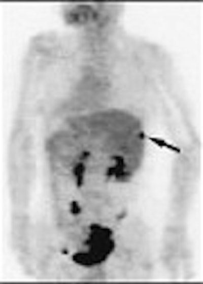
A 62-year-old woman with heterogeneous cyst of the liver. Abdominal CT scan (above) shows heterogeneous cystic lesion with suspected parenchymal liver metastasis (arrow), but not peritoneal implants on liver surface. Whole-body FDG-PET (below) scan in maximum intensity projection does not indicate significant FDG uptake in liver parenchyma (thin arrows) beside hot spot at liver surface (thick arrow). Yoshida Y, Kurokawa T, Kawahara K, Tsuchida T, Okazawa H, Fujibasyashi Y, Yonekura Y, Kotsuji F, "Incremental Benefits of FDG Positron Emission Tomography over CT Alone for the Preoperative Staging of Ovarian Cancer," (AJR, January 2004, Vol.182, pp. 227-233).
 |
The overall lesion-based sensitivity was 68% with CT and FDG-PET. The specificity was 92%, accuracy 88%, PPV 65%, and NPV 93%.
In addition, the consensus evaluation with both modalities improved correlation with postoperative staging in 87% of the 15 patients. In one case, CT alone overstaged the cancer to stage IV; combining CT with FDG-PET resulted in downstaging to stage IIIC. In another case, FDG-PET overestimated the extension of tumor to the sigmoid colon because of infiltrative activity.
"Ovarian cancer is a peculiar disease in that the detection of peritoneal metastases is not by itself a contraindication to curative surgery, but this information is important if tumor debulking is being considered. Our study has shown that the addition of FDG-PET to CT improves the accuracy in staging of ovarian cancer," the authors wrote. Proper staging can determine whether some patients should receive neoadjuvant chemotherapy before surgery.
Yoshida and colleagues also acknowledged some shortcomings of their study, including the high cost of FDG-PET and the demands it places on the patient for pre-imaging fasting. With regard to CT, the standard protocol at their institution calls for 10-mm slice thickness, although 5-7-mm may increase the accuracy of CT staging alone, they wrote.
"A major problem in ovarian cancer is that a high proportion of patients are in advanced stages of the disease at the time of diagnosis, which results in a 5-year survival rate of only 46%," the authors concluded. "Accurate staging is needed to determine appropriate treatment for those who will potentially benefit from it."
By Shalmali PalAuntMinnie.com staff writer
February 11, 2004
Related Reading
Ultrasound classification of ovarian masses can aid work-up decisions, December 19, 2003
US, CT show approximate parity for diagnosing appendicitis, December 10, 2003
Ovarian vein reflux: underdiagnosed and undertreated, June 20, 2003
Height and BMI in adolescence tied to ovarian cancer risk, August 20, 2003
PET proves versatile in imaging gynecologic cancer, August 4, 2003
Copyright © 2004 AuntMinnie.com