Wide-detector-array CT holds a number of advantages for scanning the thoracic aorta, including wider anatomic coverage of the heart and aorta, less contrast use compared to traditional scanners, and lower radiation dose, according to two new studies from the Cleveland Clinic Foundation in Ohio.
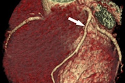
CT is the go-to modality for noninvasive evaluation of several serious diseases affecting the aorta, including atherosclerosis, aneurysm, dissection, intramural hematoma, trauma, and congenital disorders.
Using wide-area CT, radiologists can "minimize exposure time and iodinated contrast use," said Sandra Halliburton, PhD, from the Cleveland Clinic. "Wide-array-detector CT has real advantages in cardiac imaging when the aorta needs to be included due to the increased number of detector rows."
To scan the anatomy properly, radiologists need electrocardiogram (ECG) gating to avoid motion artifacts, a breath-hold to avoid respiratory artifacts, and a large field-of-view to visualize the entire thorax, and they should minimize the patient's exposure to ionizing radiation and contrast media, Halliburton said in a presentation at the International Society for Computed Tomography (ISCT) meeting in May.
Axial scanning delivers lower radiation dose
The first study examined the use of axial versus helical CT data acquisition using a wide-detector-array scanner (Brilliance iCT, Philips Healthcare, Andover, MA).
"We imaged 135 consecutive patients with suspicion of aortic disease ... with a maximum of 80-mm z-coverage per rotation," Halliburton said. Patients were scanned with either ECG-referenced helical or axial techniques depending on heart rate.
 |
| Helical and axial scanning techniques (above). According to the results for both methods (below), scan times and image quality are essentially the same, but radiation dose is dramatically lower when the axial scanning technique is used. All images courtesy of Sandra Halliburton, PhD. |
 |
"We found that we were able to image the vast majority [122/135] of our patients with axial images, [though] obviously we had a significantly higher heart rate in the helical group compared to the axial group," Halliburton said.
But with axial imaging, the radiation doses were lower, averaging 3.7 mSv versus 8.4 mSv for the helical scanning group.
"For a 280-mm scan length with gated imaging, that's a pretty good accomplishment," she said. Moreover, the mean image quality score for axial imaging trended slightly better, at 4.2 compared to 4.0 achieved with helical scanning.
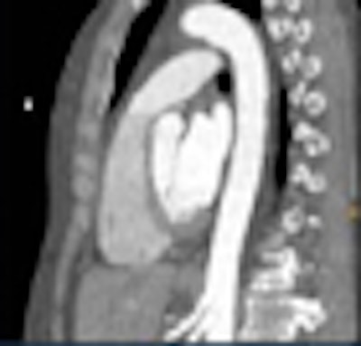
 |
| Axial scan of patient with thoracic aortic aneurysm seen on capital (left) and axial (right) views. Some photon starvation can be seen in the shoulder region, a common limitation of the axial scanning technique. |
Low tube voltage
A second Cleveland Clinic study probed the results of low-tube-voltage scanning in 43 consecutive patients with suspected aortic disease. Once again the study used ECG-triggered axial scanning techniques on the Philips Brilliance iCT wide-detector-array CT scanner, Halliburton said.
X-ray parameters (both tube voltage and tube current) were selected according to patient size -- 100 or 120 kV, and tube current time product of approximately 84 to 90 mAs.
"We felt we could image the majority of patients [37/43] with the 100-kVp technique," she said. Patients scanned with the reduced-dose protocol had a lower body mass index than those in the other group, but noise was not significantly different between the groups. Image quality remained high, while the dose was lower still compared to the previous study.
"We were able to achieve 2.7 mSv for about a 280-mm scan range," Halliburton said. "Image quality was not significantly different between the groups, and noise was not significant because of how we selected our mAs and kV," she said.
 |
| Reducing the tube current from 120 kV to 100 kV in all but the largest patients reduced radiation dose for the exam to a mean 2.7 mSv. |
Unfortunately, photo starvation was even more pronounced with the reduced parameters in axial imaging.
"We were not surprised to have even more difficulty with axial imaging through the shoulders when we lowered our tube voltage and really compared our ability to inspect the arteries," she said. "But our savior may be iterative reconstruction."
 |
| Photon starvation in the shoulder region was even more pronounced when the mAs and kV were reduced in the second study; fortunately, iterative reconstruction brought out a large amount of image detail. |
 |
Using iterative reconstruction, "we cleaned things up a little bit through the body," Halliburton said. "What's impressive is the way we were able to clean things up through the shoulders. I think the use of iterative reconstruction for these really low-dose techniques is enabling us to deal with the shoulder issue."
Wide-detector-array CT permits ECG-referenced axial imaging in the majority of patients with comparable breath-hold times as helical scanning, but it has reduced radiation dose compared to ECG-referenced helical imaging, she concluded.
In addition, imaging of the aorta can also be performed with further radiation dose reductions in some patients, using a tube current of 100 kVp, she said. Iterative reconstruction holds promise for further reducing radiation dose and improving photon starvation artifacts associated with lose-dose axial imaging, Halliburton concluded.
By Eric Barnes
AuntMinnie.com staff writer
July 7, 2010
Related Reading
F-18 NaF PET/CT can help with atherosclerotic plaque detection, June 11, 2010
Cardiac CAD safely rules out coronary artery disease, June 1, 2010
FDG-PET uptake linked to risk of acute aortic dissection, April 30, 2010
Prospective gating cuts CT aortography dose, September 22, 2009
MDCT highly sensitive for aortic dissection, March 29, 2006
Copyright © 2010 AuntMinnie.com