New concentrated contrast agents not only improve enhancement of focal liver lesions, they could make imaging safer by reducing contrast volume and flow rates, researchers in France and Germany have concluded.
The double-blind randomized parallel group study was conducted at four centers in Europe. The cohort of 151 patients included 91 with a diagnosis of abdominal tumors, and 60 patients suspected of having liver lesions, according to Dr. Renate Hammerstingl from the University of Frankfurt University Hospital in Germany, who presented the findings at the 2005 European Congress of Radiology (ECR) in Vienna.
"The most important factor for imaging focal liver lesions is contrast, and this is influenced by the administration of iodine, on the one hand by the maximum amount of iodine administration, and on the other hand the iodine delivery rate," she said. "Keeping in mind that MDCT is now much faster than the contrast distribution, we have to modify our protocols. We can increase the injection flow rate or use higher-concentration contrast medium."
The researchers chose the latter for their phase IV study, aiming to optimize iodine concentration on contrast-enhanced MDCT of liver tumors in pretherapeutic staging.
After randomizing the patients into one of three groups, Iomeprol (Bracco, Milan, Italy) was administered intravenously in one of three concentrations (300, 350, and 400 mg of iodine/mL). The overall iodine dose of 36 g was equal within the patient groups, pairing the lowest contrast volumes (range 40-80 mL) with the highest-concentration boluses (range 370-400 I/mgL).
 |
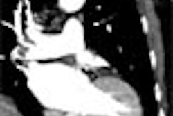
| In the hepatic arterial phase (left), a hypervascularized hepatocellular carcinoma lesion in the right liver lobe segment 7/8 (black arrow) and a second in the left liver lobe were clearly depicted. Venous-phase imaging (right) showed a decrease of signal in the small node in segment 7/8 (black arrow) and isodense contrast of the second lesion in segment 3 (white arrow). Using contrast-enhanced (Iomeprol 400 IV) MDCT, the HCC nodule (white arrow) was best seen in arterial phase, whereas the venous phase missed the lesion. Images courtesy of Dr. Renate Hammerstingl. |
"We used bolus injection by automatic power injector and a CareBolus technique," Hammerstingl said. The injection rate was 4 mL/sec for the arterial phase, and 2 mL/sec for portal-venous phase scanning, using a thin-section protocol on a four-detector MDCT scanner.
All patients underwent biphasic contrast-enhanced CT following a scan delay of 30 seconds (arterial phase) and 60 seconds (venous phase). Images were acquired using collimation of 4 x 1 mm (arterial phase) and 4 x 2.5 mm (portal-venous phase) at 160-180 mAs, 120 kVp, and rotation time of 0.5 seconds. Table feed/gantry rotation was 6 mm (arterial phase) and 10 mm (venous phase).
The primary variable for statistical analysis of contrast enhancement was the absolute contrast between liver lesions and the surrounding tissues, she said. In addition, the group analyzed contrast density of tissue of normal liver, abdominal aorta, portal-vein lesions, and surrounding lesions by summary statistics and an exploratory Kruskal-Wallis test. Image quality was considered subjectively.
 |
| Chart shows contrast density of normal liver tissue in the arterial phase. There was a statistically significant increase of density of normal liver parenchyma in the arterial phase of imaging using the highest concentration of Iomeprol (p = 0.012). The ANOVA test procedure was used for statistical analysis. Chart courtesy of Dr. Renate Hammerstingl. |
The results showed that the contrast density of normal liver parenchyma was 61.3, 64.8, and 72.7 HU for Iomeprol 300, 350, and 400 (p = 0.01), respectively, in the arterial phase. "There was a significant trend toward improved contrast using higher concentration in the arterial-phase imaging," Hammerstingl said.
In addition, the median contrast density of tissue surrounding lesions tended to be higher for Iomeprol 400 than for the other groups, both at the arterial (74 HU versus 64 and 66 HU) and portal-venous phases (113 HU versus 99 and 102 HU).
Higher contrasts of Iomeprol 400 were also measured versus the 300 and 350 formulas for subgroups of patients with certain lesions such as hepatocellular carcinoma (45, 48, and 71 HU) and cholangiocarcinoma (64, 65, and 84 HU). There was a trend toward higher densities in hypervascular lesions, but it did not reach statistical significance, Hammerstingl said.
 |
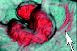
| Using high-concentration contrast, medium tumor-feeding vessels and a hypervascularized rim of the carcinoma were depicted in arterial-phase images (left). Venous phase (right) demonstrated a hypodense lesion with moderate enhancement of the periphery. Inner vascular perfusion was visualized to a high degree. Images courtesy of Dr. Renate Hammerstingl. |
"Here, an example of a patient suffering from cholangiocarcinomas," she said displaying a slide. "Note that the feeder tumor is excellently depicted in arterial-phase imaging, as well as peripheral enhancement using the high concentration, and using thinner sections, the inner architecture of the tumor is well depicted."
"Iomeprol 400 led to higher contrast density for normal liver tissue in the arterial phase of enhancement, with a maximum contrast between lesions and surrounding tissue, which was highest for Iomeprol 400, although it was not statistically significant, and we had far higher contrast for hypervascular lesions," Hammerstingl said. "With the use of higher-concentration contrast medium, you can reduce the total administered volume and you can reduce the flow rate, and that will have a convenient protocol for routine administration."
An audience member asked why the group would want to overload patients with iodine if liver lesions can generally be seen with lower concentrations. Hammerstingl noted that iodine dose is kept constant in the technique, and that previous studies have shown that the rate of adverse events drops when contrast volume and injection rates are reduced.
"What is important for hypervascular liver disease is to have a very good arterial phase, and if you use the high-concentration contrast medium with the higher iodine delivery rate within the arterial phase, that will (achieve) better contrast of the vessels of the arterial feeders toward the liver lesion itself and the liver parenchyma," she added.
Another attendee noted that late-phase CT imaging, anywhere from five to 10 minutes postcontrast administration, has been shown to improve the depiction of cholangiocarcinoma. While acknowledging those results, Hammerstingl said her group's four-detector MDCT protocol also provides excellent visualization of such lesions, and the results have improved even further using the facility's new 16-detector-row scanner.
By Eric Barnes
AuntMinnie.com staff writer
May 16, 2005
Related Reading
CT tracks renal cancer recurrence after surgery, January 20, 2005
Early arterial phase best for HCC detection, December 28, 2004
CT tracks liver involvement in HTT, February 20, 2004
For hypovascular liver tumors, late-phase CT is the way to go, August 18, 2003
All-in-one CT protocol improves evaluation of potential living liver donors, August 12, 2002
Copyright © 2005 AuntMinnie.com