A New York City firefighter, who was one of the first to arrive at the World Trade Center (WTC) following the September 11, 2001 terrorist attacks, sustained a serious case of dust-related pneumonia. Acute eosinophilic pneumonia generally presents with no evidence of infection, and can affect people with no prior history of asthma or atopic illness; imaging at the time of his hospital admission helped pinpoint the problem.
Dr. William Rom and colleagues from multiple departments of the New York University School of Medicine published their case report on the firefighter in the latest issue of the American Journal of Respiratory and Critical Care Medicine. In addition to NYU, Rom’s co-authors were from the Albert Einstein College of Medicine in New York City; the New York City Fire Department Bureau of Health Services; and Duke University Medical Center in Durham, NC.
"Acute eosinophilic pneumonia has been described as an acute febrile illness with severe hypoxemia, diffuse pulmonary infiltrates, and an increase in bronchoalevolar lavage eosinophils," the authors explained. "It is a reversible cause of noninfectious respiratory failure. The condition is idiopathic, but there have been tantalizing clues that point toward environmental dust exposure" (AJRCCM, September 15, 2002, Vol.166: 6, pp. 797-800).
The fireman worked at the WTC site for two weeks straight, 16 hours a day, inhaling large amounts of smoke and dust. He did not use respiratory protection for most of his time at the site, and eventually developed a cough that produced a "blackish sputum." He was admitted to Bellevue Hospital’s intensive care unit on September 24 with numerous symptoms including fatigue, fever, dry cough, and anterior pleuritic chest discomfort, the report stated.
The patient underwent a chest x-ray followed by a non-contrast CT scan. The latter was performed on a HiSpeed CT/1 helical scanner (GE Medical Systems, Waukesha, WI). Images were obtained at 10-mm intervals, with a slice thickness of 1-mm, from the thoracic inlet through the upper abdomen at inspiration. While the chest x-ray was standard procedure, that exam depicted "findings that were suspicious of an interstitial pneumonia that could be more accurately evaluated with the CT scan," Rom wrote in an e-mail to AuntMinnie.com.
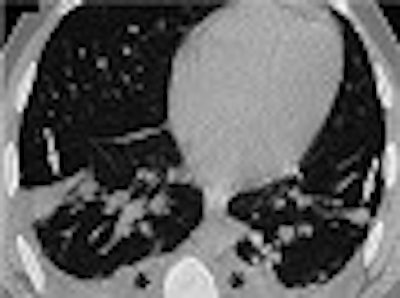
"Chest radiograph and CT scan showed patchy ground glass opacifications, thickening or respiratory airways, and bilateral pleural effusions," the authors wrote. A mineralogic analysis of the fluid from the patient’s bronchoalevolar lavage (BAL) identified 305 fibers per million alveolar macrophages. Other substances found in the patient’s lungs were fly ash, degraded glass, and amosite asbestos fibers.
 |
| On this chest CT of the 38-year-old firefighter, patchy areas of consolidation (arrows) are depicted on a 1-mm thick section at the base of the lungs. The asterisks indicate bronchial wall thickening and small pleural effusions. Image courtesy of Dr. William Rom. |
A follow-up CT scan was performed 9 days after the firefighter was treated with corticosteroids. By then, the patients pulmonary function tests improved. The second CT scan showed near complete resolution. Another follow-up three weeks after discharge indicated the disappearance of the pleural effusions, Rom said.
This firefighter was one of many to suffer from a condition that has been dubbed "World Trade Center cough." According to an article in the New England Journal of Medicine, 332 of the firefighters in the WTC rescue effort suffered from some kind of persistent cough and severe respiratory symptoms. There were other firefighters who did not suffer from WTC cough, yet still had some degree of respiratory damage. The goal of this study was to evaluate the clinical features in both of these sets of patients.
All patients underwent posterioanterior chest radiographs. These were read along with baseline x-rays taken 1-2 years before September 11 as part of the firemen’s routine healthcare protocol. High-resolution CT exams of the chest also were performed.
In 96% of the firefighters with WTC cough, chest x-rays were unchanged from baseline, the study stated. Of the 78 subjects with normal findings on chest x-ray who then underwent CT, 28% had no abnormalities. Air trapping was seen in 51% of these patients, and 30% also had bronchial wall thickening. Isolated parenchymal findings or parenchymal findings in combination with airway abnormalities were identified in 10% of the firemen.
"The clinical and physiological findings in patients with WTC cough and the airway-responsiveness findings in the cohort of firefighters who were exposed but in whom the cough did not develop demonstrate that there was a clinically significant respiratory exposure," the authors wrote. "(Ninety-five) percent had symptoms of dyspnea, 87% had gastroesophageal reflux disease, and 54% had nasal congestion. Of those tested before treatment of World Trade Center cough, 63% of firefighters had a response to a bronchodilator and 24% had bronchial hyperreactivity" (NEJM, September 12, 2002, Vol.347:11, pp. 807-815).
Although the intensity of exposure at the WTC went far beyond the ordinary, these studies emphasize the importance of protective devices for rescue workers, wrote Dr. William Beckett in an accompanying AJRCCM editorial. Beckett is with the department of environmental medicine at the University of Rochester School of Medicine and Dentistry in New York.
"Observers in New York noted that...many experienced rescuers did not use (protective devices) consistently, foregoing their own safety in the effort to work more effectively," Beckett said. "The individual respirator, like the seat belt, is effective only when actively used. Perhaps the (WTC) disaster can spur engineers and material scientists to find new ways to protect the lungs of those who risk their lives to save others."
By Shalmali PalAuntMinnie.com staff writer
September 25, 2002
Related Reading
Half-dose chest CT produces acceptable image quality, August 5, 2002
Flat-panel DR shrinks chest x-ray dose, May 27, 2002
Chest CT pegs inhalation anthrax at Virginia facility, October 30, 2001
Copyright © 2002 AuntMinnie.com