One of the most serious conditions associated with the dog days of summer is heat stroke. The National Oceanic and Atmospheric Administration estimates as many as 200 people die a year in the U.S. from heat-related disorders, particularly during heat waves. From 1979-1999, excessive heat exposure caused 8,015 deaths in the U.S., according to the Centers for Disease Control and Prevention's Extreme Heat Web site.
Heat stroke occurs when the body is unable to regulate its temperature, which then rises rapidly. The sweating mechanism fails and the body cannot cool down. The elderly are especially prone to heat-related health problems for a variety of reasons, including a naturally occurring reduced ability to sweat, decreased mobility, and, in some cases, medications that thwart thermoregulation.
An interdisciplinary team from Israel found that CT scans could be used to track the changes in the central nervous system (CNS) of heat-stroke victims. They reported their findings in the European Journal of Radiology.
"The CNS is very sensitive to elevation of body temperature: its dysfunction following elevated body temperature leads to coma and brain death, not infrequently causing patient demise. We describe the pattern of CNS changes as detected by brain CT," wrote Dr. Oded Szold and colleagues. Szold, along with Drs. Ron Ben Abraham, Philippe Biderman, and Patrick Sorkine are from the general intensive care unit (ICU) at Tel Aviv Sourasky Medical Center, Tel Aviv University. Drs. Irith Reider-Groswasser, Galit Aviram, and Yoram Segev are from the medical center’s department of radiology (EJR, July 2002, Vol.43:1, pp.1-5).
For this study, five males and one female with a mean age of 46 years were admitted to the ICU during a 1999 summer heat wave in Israel. They all fulfilled the criteria for heat stroke, including a core body temperature of over 40° C, altered mental status, and dry skin.
"All six arrived in a deep comatose state accompanied by areflexia and the absence of brain stem reflexes," they wrote.
"The main purpose of the CT imaging is to rule out any treatable CNS problem, such as bleeding (due to coagulopathy)," added Szold in an e-mail to AuntMinnie.com.
Noncontrast CT exams were performed, within four days of admission, on Elscint scanners. Adjacent slices of 2.5 mm were acquired of the posterior fossa and 10-mm slices of the rest of the brain (imaging matrix = 326 x 326 pixels). CT findings were assessed using the following parameters:
- Presence or absence of fresh bleeding.
- Focal lesions.
- Ventricular size.
- Sulcal size.
- Cisternal size.
- Gray and white matter discriminability (GWMD).
GWMD was graded on a scale of 0-3, with 0 indicating a normal pattern and 3 representing no white-matter digitations.
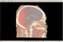
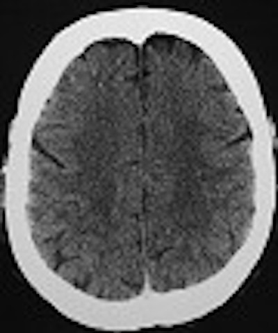
According to the results, "all six patients demonstrated severe loss (grade 2-3) of the GWMD. None suffered from CNS bleed or displacement of anatomical structures. The difference on CT between the gray and the white matter is thought to be due to the higher content of water and lower content of lipids in the gray matter. This results in higher oxygen and lower carbon concentrations in the gray matter, causing increased photoelectric absorption," they said.
 |
Axial cuts at the levels of the lateral ventricles and centrum semiovale (mAs 335, kv 120). A loss of GWMD is shown in a 51-year-old comatose patient with exertional HS in comparison to good GWMD in a 50-year-old patient, below. Images courtesy of Dr. Oded Szold.
 |
Two patients died of septic shock and multiorgan dysfunction syndrome while in the hospital. One patient, on the fourth day in the ICU, responded to deep pain stimuli. He was fully conscious 11 days after admission.
Of the three remaining patients, one was fully awake within 48 hours; one demonstrated slow neurological improvement and was back to his cognitive and emotional baseline within 8 weeks; the last patient remained in a vegetative state at the six-month follow-up. As of August 2002, Szold’s group was trying to track down this patient for additional imaging studies to determine if further GWMD had taken place.
CT scans of heat-stroke patients' altered consciousness are now done routinely at his institution, although only after the patient’s body temperature has been cooled to 38.5° C, Szold said. Deciphering whether GWMD is merely an indicator of early brain-tissue damage in heat stroke, or whether it can predict the patient’s clinical course, will require larger studies, including spectroscopic analysis of the gray-white junction, the group concluded.
By Shalmali PalAuntMinnie.com staff writer
August 14, 2002
Related Reading
MRI trains an eye on orbital Lyme disease, June 10, 2002
Frontal gray matter structure highly influenced by genetics, November 6, 2001
Copyright © 2002 AuntMinnie.com