Introduction
Along with the bobsled and the skeleton sled, the Winter Olympics sport of luge involves a fast ride down a tortuous man-made ice course. The sled has no brakes, and as luge athletes steer down the course they can reach speeds of 80 mph or more. Yet despite the harrowing course and the high speeds, luge appears to be relatively safe, with injury rates comparable to those of recreational Alpine skiing (American Journal of Sports Medicine, July-August 1997, Vol. 25:4, pp. 508-513).
Clinical history
Male, 31, with wrist pain after sustaining an injury while paddling (propelling the sled with one's hands while wearing spiked gloves).
Physical exam and radiographic findings
The patient had point tenderness over the medial volar portion of the distal right wrist. He reported that the wrist was most painful with the palmar flexion against resistance that was necessary to properly propel the sled.
He also reported a similar injury at the end of the previous year’s season, but sought no medical attention at the time. The initial radiographs obtained seven days after the original injury were negative. Due to the prominent clinical symptoms, the presence of point tenderness, and the mechanism of injury, an MRI was obtained for further evaluation.
Diagnosis
Old ununited fracture of the hook of the hamate.
Discussion
Most of the current information regarding luge-related injuries was obtained from athlete injury and illness report forms at the U.S. Training Center Sports Medicine Clinic in Lake Placid, NY. The risk of sustaining an injury was 0.39 injuries per person per year. The risk of injury causing the loss of more than one day of practice was 0.04 injuries per person per year.
Thus, an injury is sustained by one out of three luge athletes for every 55 runs on the track. This is comparable to injury rates seen in recreational skiing (American Journal of Sports Medicine, July-August 1997, Vol. 25:4, pp. 508-513).
The most common types of injuries were contusions (51%) and strains (27%). Fractures accounted for 3% of the total number of injuries, and crashes were responsible for most of the injuries seen at the clinic (American Journal of Sports Medicine, July-August 1997, Vol. 25:4, pp. 508-513). Fractures of the hamate are relatively unusual, representing only 2%-4% of fractures of the carpal bones (Hand Clinics, February 1987, Vol.3:1, pp.149-161).
Hamate fractures may occur anywhere within the bone itself, but fractures of the hook of the hamate are especially important due to the complications seen in this type of fracture (osteonecrosis, injuries to the surrounding tendons, and nonunion (Diagnosis of Bone and Joint Disorders, Harcourt Health Sciences, Philadelphia, 1995).
Hook of the hamate fractures are generally thought to have two mechanisms, resulting either from a fall on the wrist with the hand in dorsiflexion, or by direct trauma. The latter mechanism is the one most commonly encountered in athletic injuries, and is usually seen in sports that involve clubs or bats (golf, baseball).
Clinical diagnosis is often difficult, as it is an uncommon type of fracture and can present with atypical symptoms, relating to involvement of surrounding tendons (the extensor digiti minimi or the extensor carpi ulnaris) or nerves (the ulnar or median nerve). The difficulty is compounded by the fact that conventional x-rays are usually negative, and adequate plain-film evaluation requires the use of carpal tunnel views for an optimal view of the hook of the hamate.
Cross-sectional imaging is frequently needed to either confirm or exclude the possibility of a fracture of the hamate. Axial T1 and/or axial proton-density MR images best display the hook of the hamate and are the preferred imaging sequences when an injury to this region is suspected.
T2-weighted images with fat suppression are also very helpful in demonstrating the bone marrow and soft-tissue edema that is associated with an acute fracture. Coronal images, often obtained in MR imaging of the wrist, are limited in their ability to display most fractures of the hook of the hamate, due to the coronal orientation of most of these fractures.
Thin-section helical CT is the preferred method of fracture detection. Thin sections (1-1.5 mm) can accurately identify the presence of a fracture, its extent, and any involvement with the surrounding articular surfaces.
 |
| The axial, proton density MR image above demonstrates an oblique fracture through the base of the hook of the hamate (black arrow). The sclerosis at the margins of the picture is consistent with the clinical report of old trauma. |
 |
| Above, an axial T2-weighted MR image shows the oblique fracture through the base of the hook of the hamate with intervening fluid (black arrow). The increased T2 signal, indicative of edema and inflammation (white arrowheads) corresponded to the location of the patient's pain. |
 |
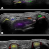
| Coronal MR images illustrate the difficulty in visualizing fractures of the hook of the hamate. These fractures (black arrows) are in the coronal plane, and are therefore difficult to visualize on both the T1-weighted (right) and gradient echo (left) images. Images courtesy of Dr. Douglas P. Beall. |
Conclusion
Although fractures are not common in luge, they should not be overlooked as a cause of morbidity and loss of peak performance. Fractures of the hook of the hamate can have a varied clinical presentation and can be difficult to diagnose, both clinically and radiographically. Cross-sectional imaging should be strongly considered in any patient suspected of having a hamate fracture.
By Dr. Douglas P. BeallAuntMinnie.com contributing writer
February 22, 2002
Dr. Beall is a staff radiologist in the musculoskeletal division, department of radiology at Wilford Hall Medical Center, Lackland Air Force Base, TX. He also is an assistant professor of radiology, department of radiology and nuclear medicine, Uniformed Services Health University in Bethesda, MD.
The opinions and assertions herein are the private views of the authors and are not to be construed as official or as reflecting the view of the Department of the U.S. Air Force, U.S. Department of Defense, or the U.S. government.
Related Reading
Luge brings contusions, concussions, fractures, February 12, 2002
Copyright © 2002 AuntMinnie.com