Patients who present to the emergency room with hemorrhaging caused by pelvic fractures can be challenging trauma cases for radiologists. Plain radiographs are useful for detecting hemorrhagic sites and for determining femoral access sites for embolization in hemodynamically unstable patients, according to Japanese researchers.
Dr. Tetsu Niwa and colleagues from the department of radiology at Yokohama City University School of Medicine and the critical and emergency care center at Yokohama City University Medical Center have developed protocols for the initial management of unstable pelvic fractures as a result of their research, which was presented in a poster presentation at the 2002 RSNA conference in Chicago.
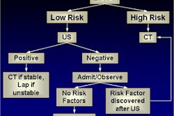
The group noted that intravenous contrast-enhanced CT of the pelvis is highly accurate in determining the presence or absence of an ongoing pelvic hemorrhage in hemodynamically stable patients. However, unstable pelvic fractures are a significant cause of morbidity and mortality in patients with multiple injuries.
"In hemodynamically unstable patients with pelvic fractures, a CT scan for the evaluation of pelvic fractures has been controversial and impractical in some hospitals because it may be time-consuming and transport can be difficult and considerably aggravate the hemorrhaging," they wrote.
Use of a prompt and precise embolization technique is critical due to the danger of death by exsanguination resulting from a delay in hemostasis. Open surgical exploration of arterial bleeding fails to gain access to the femoral arteries, according to the researchers.
The group used a modified Bassam protocol, first published in the American Journal of Surgery for its initial management of unstable pelvic fractures. For anterior fractures, they conduct a primary external fixation as well as angiographic embolization for a continued hemorrhage. For posterior fractures, the group conducts a primary angiographic embolization, then performs an external fixation for a continued hemorrhage (American Journal of Surgery, September1998, vol. 64:9, pp. 862-867).
 |
| Image and caption provided courtesy of Dr. Tetsu Niwa and colleagues, Yokohama City University School of Medicine and Medical Center. |
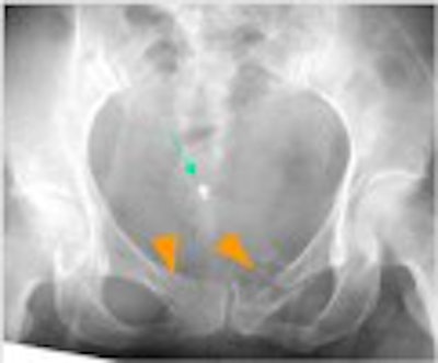
The researchers noted that plain radiographs provide the radiologist with the most rapidly available images for hemodynamically unstable patients. However, before therapeutic procedures can be inaugurated, a careful interpretation of the radiographs and clinical findings is required.
"Plain radiographs can indicate hemorrhagic sites in the anterior of the pelvis with high sensitivity. Posterior fractures in the pelvis should be interpreted carefully, because they can sometimes be difficult to identify and they associate with greater hemodynamic instability," they wrote.
 |
| Image and caption provided courtesy of Dr. Tetsu Niwa and colleagues, Yokohama City University School of Medicine and Medical Center. |
The radiologists at Yokohama City University Medical Center use plain-film radiographs to determine lateralization of the severity of the hemorrhage prior to angiographic intervention. This requires that the radiologist examine the fracture pattern, including the direction and intensity of the wounding force, to determine the appropriate femoral access sites.
 |
| Image and caption provided courtesy of Dr. Tetsu Niwa and colleagues, Yokohama City University School of Medicine and Medical Center. |
The team observed that plain radiographs in multiple trauma patients who are hemodynamically unstable may show false-positive results, particularly with the posterior segments of the pelvis. This may be due to intrapelvic structures such as colon gas, which can obstruct a lesion in the posterior division of the internal iliac artery.
Also, subtle fractures may be difficult to detect via an anteroposterior view with a plain radiograph. In addition, the peripheral branches of the anterior division of the internal iliac artery can be spastic and may not reveal an ongoing hemorrhage.
"Pelvic injury should be evaluated with the patient’s history and physical examination, as well as plain-film radiographs," the researchers wrote.
When performing an arterial embolization as the next step in stabilizing the patient, the radiologist’s interventional management is aimed at stopping massive hemorrhaging as quickly as possible. The group omits pelvic angiograms and performs selective internal iliac angiograms for mapping purposes.
 |
| Graphic provided courtesy of Dr. Tetsu Niwa and colleagues, Yokohama City University School of Medicine and Medical Center. |
The researchers then target the arterial branches at the most severe site, using a 5-French catheter that is placed into the internal iliac artery via the contralateral femoral artery. When necessary, they perform superselective catheterization of the anterior or posterior branch with a coaxial 3-French catheter.
The interventional radiologists use gelatin sponge particles to embolize anterior areas, then stabilize them with steel coil (3 mm to 5 mm diameter) or a fibered platinum microcoil embolization is performed. For posterior braches, they embolize by coil only. To prevent complications they avoid excessive embolization of arteries other than the internal iliac arteries, and try to avoid overlapping embolization whenever possible.
"Predicting arterial injury sites and determining an appropriate access site can be accomplished by the interpretation of plain-film radiographs before arterial embolization," the authors wrote. "Awareness of the pattern of pelvic fracture and their plain-film radiographic features help to perform prompt and effective intervention."
By Jonathan S. BatchelorAuntMinnie.com staff writer
May 9, 2003
Related Reading
Residents' after-hours CT interpretation shows high accuracy, March 4, 2003
Plain film x-rays not needed with CT to evaluate polytrauma spinal injuries, December 6, 2002
Pediatric ankle exam predicts need for x-rays, November 1, 2002
Emergency Imaging of the Acutely Ill or Injured Child, September 10, 2002
Education is the solution for on-call radiology, February 21, 2002
Copyright © 2003 AuntMinnie.com