A new study in Radiology examines the use of teniae coli -- three thick bundles of muscle that run along the outer longitudinal layer of the colon's muscularis externa -- as a navigation and localization tool for virtual colonoscopy (VC or CT colonography [CTC]) data.
Researchers from the National Institutes of Health (NIH) in Bethesda, MD, believe that teniae coli, as extractable features of VC datasets, can significantly improve polyp localization over current methods, potentially improving specificity as well by enabling the examination of prone and supine datasets side by side.
Their retrospective study sought to develop a circumferential localization method based on teniae coli to guide VC navigation and polyp registration.
Two main types of reference systems, both of which have drawbacks, are currently used for polyp registration at CT colonography, noted NIH authors Adam Huang, Ph.D.; Dr. Dave Roy; Dr. Ronald Summers; and their colleagues. The team included Dr. Perry Pickhardt from the University of Wisconsin at Madison, who supplied data from the U.S. Department of Defense (DoD) CTC trial (New England Journal of Medicine, December 4, 2003, Vol. 349:23, pp. 2191-2200) for use in the Radiology study.
"In one system, the colon is divided into different anatomic sections; the other system relies on the endoluminal centerline distance along the colon," the group wrote. "The sectional approach is not precise because the anatomic sections are relatively long and their boundaries are poorly defined (Radiology, December 2007, Vol. 243:2, pp. 551-560).
The centerline approach, while offering more longitudinal precision than the anatomic section approach, lacks orientation information by which the circumferential position of a lesion on the colonic wall could be ascertained.
"This drawback makes polyp registration between scans time-consuming and subject to error," they wrote.
The teniae coli, three approximately 8-mm-wide longitudinal bands of smooth muscle that extend from the cecum to the sigmoid colon, make a better potential navigation and localization tool than the others because they run the length of the colon, and their width remains fairly constant until they widen at the level of the sigmoid colon, the authors stated. The muscles aren't readily visible on 2D axial views, but can be easily seen as continuous flat bands in 3D VC endoluminal views, the investigators noted.
The study was performed on 36 VC cases representing 26 men and 10 women (mean age 59). The data were selected from the San Diego Medical Center studies that were included in the Department of Defense trial, which examined asymptomatic subjects with same-day virtual and optical colonoscopy.
The selected data were those that contained at least one colonoscopy-proven polyp 6 mm or larger, located between the cecum and the descending colon, and visible on both prone and supine datasets, the authors stated.
The subset of cases Huang et al studied contained 47 polyps, including 13 (28%) pedunculated and 34 (72%) nonpedunculated (33 sessile, one flat). Two polyps (1.0 and 4.2 cm) were cancerous, 38 were adenomatous, five were hyperplastic and two were benign, the group noted.
All subjects underwent VC following cathartic cleansing and self-administered colonic insufflation with room air, and same-day conventional colonoscopy to confirm the presence of VC-detected polyps.
In each case the tenia omentalis (TO), the most readily visible of the three teniae coli on a well-distended colonic surface, was manually extracted from the cecum to the descending colon, using software developed in-house. The researchers performed 3D visualization using proprietary software (V3D Colon, Viatronix, Stony Brook, NY).
By virtually dissecting and flattening the colon along the TO, the authors developed a localization system involving 12 grid lines to estimate the circumferential position of polyps, the authors wrote.
"The TO points were selected from the cecum through the descending colon only, because the teniae coli become difficult to distinguish anatomically in the sigmoid colon," they explained. "The manual extraction generally takes 10 to 15 minutes per colonic surface. The shortest path (graph geodesic) through the TO points was then calculated and drawn on the surface by the software."
 |
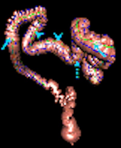
| Teniae coli and minor grid lines on anterior (above) and posterior (below) views of the 3D CT colonographic surface. The TO (solid arrows), TL (dashed arrow), and TM (arrowheads) were mapped from a 2D sheet back to the 3D surface. Figures 3a and 3b republished with permission of the Radiological Society of North America from Adam Huang, Dave A. Roy, Ronald M. Summers, Marek Franaszek, Nicholas Petrick, J. Richard Choi, and Perry J. Pickhardt, "Teniae Coli-based Circumferential Localization System for CT Colonography: Feasibility Study" Radiology 2007; 243: 551-560. |
 |
Based on the location of the extracted TO, the software inferred the positions of the other two teniae coli, the tenia libera (TL) and tenia mesocolica (TM), assuming the latter two muscles were equidistant from the TO.
From the extracted TO "we derived a centerline of the unflattened colon as the average 3D position of the teniae coli," Huang et al wrote. "From the centerline and the TO, we defined a local coordinate system by letting the tangent of the centerline be the z direction and the vector pointing to the TO to be the y direction."
The synchronous navigation system prototype was based on side-by-side VC view panels for prone and supine datasets, and applied to all datasets. "For each polyp, a signed circumferential position difference between scans was computed by subtracting the supine scan position of the polyp from the prone scan position," the team wrote.
Reconstructed colon surfaces were color-coded: in pink to represent normal mucosa, yellow-orange to represent polyp candidates.
The researchers were able to extract the TO manually in all but two (97%) of the 72 colonic surfaces. And the software-generated TM and TL paths -- inferred as equidistant from the TO path -- corresponded approximately to the locations the researchers visualized in the datasets.
Still, 8% of 72 colonic surfaces had erroneous TL coordinates in a small portion of the inferred TL, owing to various causes including leakage into the small bowel in three cases, holes on segmented surfaces (one case) and polyps touching different colonic sections in two cases. Ninety-six percent of the 47 polyps were within the range of extractable TO predicted from both scanned positions. Two other polyps were located in a poorly distended distal descending colon where the TO could not be traced.
"By orienting and positioning the virtual cameras with use of the new localization system, synchronized prone and supine navigation was achieved," the authors reported. "Our study results indicate that a polyp found on one (prone or supine) scan would most likely (with 95% confidence) be found within ± 6.1 cm on the complementary scan." And since radiologists are often able to characterize the morphology of polyps seen on VC data, this knowledge can help define the search area, they added.
Teniae coli are "recognizable intrinsic landmarks" that can be extracted from CTC data by finding the TO path and flattening the colon along its path, the group concluded. In addition to increasing the accuracy of polyp localization and lesion matching between prone and supine datasets, the method could be helpful for orienting 2D scans between acquisitions and synchronizing current and prior scans.
The main limitations of the study are that TO extraction must be performed manually, and is user-dependent, thus potentially introducing errors, the authors noted. And the method's utility does not extend to the distal colon, where the TO is difficult to distinguish anatomically.
In nearly all cases, however, teniae coli-based navigation "complements the conventional centerline approach for virtual colon navigation and polyp registration in CT colonography," they wrote.
By Eric BarnesAuntMinnie.com staff writer
January 2, 2008
Related Reading
VC measurements more accurate than optical colonoscopy, May 30, 2007
CAD will be key to VC screening, September 13, 2006
Colon muscles do heavy lifting in polyp positioning, October 19, 2005
Colonoscopy often inaccurate in localizing colorectal cancer, October 18, 2005
VC path-planning technique maximizes observable mucosa, August 30, 2005
Copyright © 2008 AuntMinnie.com