Omental infarction is an uncommon cause of severe abdominal pain in both children and adults, especially men. Some cases are associated with prior surgery, trauma, or omental torsion, while others may be caused by anomalies in the arteries supplying the omentum, or the kinking of veins associated with increased intra-abdominal pressure.
Omental infarction (OI) rarely requires surgery, but can it be mistaken for other conditions, such as acute appendicitis, that do need intervention and quickly. Thus it is the differentials that make correct diagnosis critical, and that's where thin-section CT comes in.
A five-year series from the University of Pittsburgh Medical Center in Pennsylvania aimed to shed light on the subject by assessing the prevalence, presentation, and type of OI found in both children and adults. The research, presented at the 2006 RSNA meeting in Chicago, found predictable patterns and presentations that could speed diagnosis and decision-making, and perhaps avoid unnecessary intervention.
"Omental infarction is an uncommon but increasingly recognized cause of acute abdominal pain," said Dr. Camilo Borrero, of the University of Pittsburgh Medical Center and Children's Hospital of Pittsburgh, in his presentation. "It's been noted in the surgical literature for almost 100 years, but has come to the increasing attention of radiology in the past 15. This is thought to be due to the increased use of CT, as well as the obesity epidemic," which is thought to be a risk factor for OI.
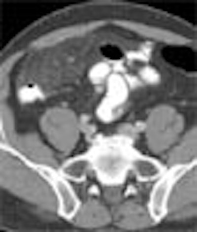
 |
| Primary OI in a 51-year-old man. Note torsion of vessels, and heterogeneous fatty mass adjacent to ascending colon. All images courtesy of Dr. Camilo Borrero. |
Most OI patients are adults, who present with right-sided abdominal pain -- usually the right lower quadrant, he said. OI can simulate conditions such as appendicitis and diverticulitis, but OI doesn't require surgery.
"The purpose of our study was to compare primary and secondary forms of omental infarction, including the demographics and location," Borrero said. "In particular, we looked at the appearance -- usually a fatty mass -- as well as the presence or absence of thickening in the adjacent bowel wall," he said. Torsion, or twisting of the omental vessels, may or may not be present.
Borrero, along with Drs. Omar Almusa, Manuel Meza, and Michael Federle, retrospectively examined CT images from 65 OI patients over a five-year period, including adults and children, and patients with both with primary and secondary forms of the disorder.
 |
| Primary OI in an 11-year-old boy, without torsion. |
Primary means there was no prior surgical or inflammatory process known to cause omental infarction, he said, while secondary OI was used to refer to patients with prior surgery, inflammation, hernia, etc. as a likely etiology.
Of the 65 patients studied, the researchers found primary OI in 21 patients, including nine children and 12 adults. The children ranged in age from 4-18 years (mean 9) and the adults were age 20-63, with a mean age of 44 years.
"This goes along with the literature: most patients were male -- 16 of 21 -- and almost all patients (20/21) had pain and tenderness in the right side of the abdomen" (82% right lower quadrant, 15% right upper quadrant), Borrero said. Finally, five of 21 patients had torsion of the vascular pedicle, and the other 16 had a fatty mass without torsion.
 |
| Secondary OI in a patient with prior sigmoid resection for diverticulitis. |
The researchers found 44 cases of secondary OI, including three children and 41 adults (22 men, 22 women; ages 1-87, mean age 49 years).
But unlike the primary OI patients, complaints of pain and tenderness in the secondary OI patients were distributed throughout all quadrants of the abdomen -- only 30% were on the right side, Borrero said. And the site of symptoms corresponded to the site of prior surgery, hernia, or inflammatory process.
There was prior abdominal surgery in 33 patients, including intestinal (n = 14), appendix (n = 5), pancreas (n = 5), stomach (n = 1), liver (n = 3), gallbladder (n = 4), and ventriculoperitoneal (VP) shunt removal (n = 1).
Five patients had hernias, including ventral (n = 2), umbilical (n = 2), and inguinal (n = 1). The etiology was inflammatory in six cases, including appendicitis (n = 2), pancreatitis (n = 1), ulcerative colitis (n = 1), Crohn's disease (n = 1), and ischemic colitis (n = 1).
Of the 41 primary OI cases, seven patients (five with torsion and two nontorsion) underwent surgery. The diagnosis was missed by the radiologist in one primary case, the diagnosis was "not believed by the surgeon" in four primary cases, and worsening pain led to surgery in two primary cases, Borrero said.
 |
| Secondary OI in an 11-year-old boy, with torsion from ruptured appendicitis. |
As for their disposition, five of the secondary OI patients underwent subsequent surgery, including three patients with hernias, and two cases of perforated appendicitis. The remaining OI patients had no surgery and all but two had a favorable clinical course, he said. The exceptions were an abscess in secondary OI (drained percutaneously), and a hemorrhage in primary OI.
"Omental infarction can occur in both adults and in children (40% primary and 7% secondary), although it tends to occur more in adults. Also when it occurs in adults, it tends, based on our series, to be in the secondary form (60% primary and 93% secondary)," Borrero said.
Primary OI is usually adjacent to the ascending colon in both children and adults, he said, while secondary OI occurs adjacent to the underlying cause, such as prior surgery, hernia, or inflammatory focus, he said.
"Lastly and most importantly, I think, high resolution of CT allows us to distinguish and properly characterize these infarcts, and avoid unnecessary surgical intervention."
By Eric Barnes
AuntMinnie.com staff writer
January 3, 2007
Related Reading
MDCT finds cause of acute abdomen more often with MPR, November 29, 2006
The more CT detectors, the more MPRs help, March 22, 2006
Scores of detector rows bring opportunities, challenges to CT imaging, April 8, 2005
MDCT yields reliable diagnosis of pancreas divisum, April 14, 2005
Copyright © 2007 AuntMinnie.com