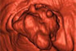
ORLANDO, FL - Three-dimensional visualization with PET/CT may soon be expanded to virtual bronchoscopy, providing a new tool for diagnosis, treatment planning, and interventional guidance, according to a multidisciplinary group from Stanford University in California.
"While CT-based virtual fly-through technology is available in the clinic, this modality lacks the sensitivity and molecular specificity required for routine use that PET/CT-based fly-through stands to offer," the authors wrote.
A team from the departments of bioengineering, radiology, cardiothoracic surgery, nuclear medicine, and molecular imaging at Stanford published a poster presentation demonstrating the utility of PET/CT-based virtual bronchoscopy at the 2006 Academy of Molecular Imaging (AMI) scientific meeting this week.
The group created an anatomically accurate plastinated porcine heart lung phantom to simulate airways and peripheral structures. They then positioned flat and polyploid-shaped plastic capsules, sized between 4 mm to 10 mm, with varying levels of F-18, 0.5 µCi to 2 µCi, into the trachea and bronchial tree, according to the authors. Sequential images were acquired with a Discovery LS PET/CT system (GE Healthcare, Chalfont St. Giles, U.K.).
The CT protocol utilized helical imaging, a 2.5-mm slice thickness, a pitch of 0.75 to 1, and 100 mAs and kVp. Reconstruction was filtered back-projection. PET images were acquired in 2D imaging at five minutes per bed position, with ordered-subsets expectation maximization (OSEM) reconstruction of 5-mm slices in two iterations, the team wrote.
Virtual bronchoscopy fly-through movies and images were created by the scientists using GE's Volume Viewer Plus and custom software. The location of the capsules was validated using standard video bronchoscopy.
"Both polyploid-shaped capsules were visualized using CT virtual fly-through," the authors wrote. "However, only one of three flat capsules was visible using CT only."
The researchers reported that all five placed capsules were identified in axial PET/CT images. Furthermore, the flat lesions not identified on the CT virtual fly-through were visible using PET/CT fly-through visualization.
The group said its future work will focus on several key areas of the technology, including simulating other realistic conditions such as variation in lesion activity, locating lesions in areas adjacent or peripheral to the airways, and background activity. In addition they plan to further study optimizing the visualization parameters for PET/CT-based virtual bronchoscopy.
"More studies will be required to determine the quantitative detection threshold for different levels of background and noise," they wrote. "Ultimately, animal studies and patient data will be required to prove the clinical utility of this new and promising technology."
By Jonathan S. Batchelor
AuntMinnie.com staff writer
March 30, 2006
Related Reading
3D rendering captures SNM's Image of the Year, June 21, 2005
3D: Rendering a new era, May 2, 2005
4D ultrasound shows promise for improving fetal echocardiography, April 6, 2005
3D volume US shows promise for second-trimester fetal evaluation, March 17, 2005
FDA approves 3D CT bronchoscopy, November 15, 2004
Copyright © 2006 AuntMinnie.com


