Given the unreliability of initial radiographs in detecting scaphoid fractures, another inexpensive approach to imaging everyday wrist trauma should be welcome. But with so many other modalities available, the panoramic view may be too unusual to embrace.
Not that orthopantomography is unusual per se. It is very commonly used, of course, for dentomaxillofacial imaging. Nonetheless, it may be the rare facility that can integrate panoramic x-rays into wrist imaging as physicians from the University General Hospital in Murcia, Spain, have done.
In their latest study of this approach, the Murcia investigators report that orthopantomography detected 21% more suspected scaphoid fractures than plain radiography (American Journal of Roentgenology, January 2004, Vol. 182:1, pp. 155-159).
Their study looked retrospectively at 90 patients, ages 11 to 72, who had presented at the hospital with acute or chronic wrist trauma. All had been examined with conventional radiography in four projections (posteroanterior, lateral, oblique in pronation, and ulnar deviation) and with one or two posteroanterior panoramic projections.
Panoramic images were taken with an Orthoralix SD Ceph machine (Philips Medical Systems, Monza, Italy) using 60-64 kV, 4-6 mA and a consistent 12-second rotation time. The patients’ wrists were immobilized using a device patented by radiologist Dr. Juan Berná, lead author of the study.
 |
| Patient's wrist positioned for panoramic imaging in the Berná immobilizing device, which will be marketed by Dispositivos Médicos de Imagen of Albatera, Spain. Image couresty of Dr. Juan Berná. |
"With the patient sitting, standing, or preferably lying down, the standard posteroanterior panoramic projection was made with the forearm vertical and the hand in supination or with the wrist placed in a sagittal plane parallel to the sagittal sinus of the patient," the authors wrote. "The variant of the standard posteroanterior panoramic projection was performed using approximately 10° of rotation of the wrist toward the radial side."
  |
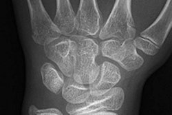
| Forty-nine-year old woman with acute trauma of the wrist. Conventional radiography and panoramic radiography were performed two weeks after injury. Conventional radiographs (above) show no evidence of fracture. |
The retrospective readings were done by two traumatologists and two radiologists with five to 25-plus years’ experience. They were given brief training on panoramic wrist x-rays as none previously reviewed such images.
The review found panoramic radiography of the wrist superior in ruling out scaphoid fractures (74%, 20/27) in patients with suspicious findings on conventional radiography. Panoramic images also revealed more cases of scaphoid fractures (21.4%, 12/56) and more cases of delayed union, nonunion, and union.
 |
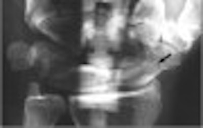
| Panoramic radiograph (above) shows fracture line (arrow, fracture type A2). Berná J, Chavarria G, Albaladejo F, Meseguer L, Pellicer A, Sánchez-Cañizares, M, Pérez-Flores, D, "Panoramic Versus Conventional Radiography of Scaphoid Fractures," (AJR, January 2004, Vol.182, pp.155-159) |
"In our hospital, for several years now, orthopedic surgeons have requested panoramic radiography whenever the findings of conventional radiography suggest a possible but doubtful diagnosis of scaphoid fracture or nonunion," the authors wrote. "We recommend this procedure as a complement to conventional radiography for the clarification of inconclusive conventional radiographic studies."
The demand for something better than just x-rays is widespread. Delayed diagnoses occur more often with wrist fractures than nearly any other traumatized area, and many surgeons recommend immediate operative intervention for displaced scaphoid fractures.
The "notorious difficulty" presented by scaphoid fractures has also provided steady fodder for investigators, with recent reports finding that bone scintigraphy, CT, MRI, and sonography are all better at detecting scaphoid fractures earlier.
Indeed, researchers from Duke University in Durham, NC, reported that an immediate MRI protocol was cost-effective at their insititution, while French researchers reported 98% accuracy with high-resolution sonography (AJR, December 2001; Vol. 177:6, pp. 1257-1263; AJR, May 2002, Vol.178:5, pp.1239-1245).
However, the American College of Radiology’s updated Appropriateness Criteria for Acute Hand and Wrist Trauma recommends MRI only if immediate confirmation or exclusion of a fracture is required, and CT only if x-rays show a scaphoid fractures and displacement or fracture age is a concern, or if repeat radiographs after cast application are negative.
Otherwise, the guidelines stand firmly behind the standard practice for suspected scaphoid fractures with normal radiographs: apply a cast and repeat the x-ray exam after 10 days. The 2001 recommendations also make no mention the panoramic option first posited by Berná and colleagues in the early 1990s.
That omission won't likely change with the planned 2006 guideline update, according to Dr. David Rubin, a musculoskeletal specialist at the Mallinckrodt Institute of Radiology in St. Louis and primary author of the ACR’s updated wrist imaging criteria.
Commenting on the Spanish study, Rubin wrote in an e-mail to AuntMinnie.com: "I don't think using Panorex examinations for scaphoid fractures will ever come into widespread use. I am concerned that the parts of the fracture that are out of the focal plane are blurred (mimicking callus formation)." He also noted that most radiology departments have multiple CT scanners and only one Panorex machine, and that panoramic imaging requires a special wrist holder.
But while Berná may address the last problem with plans to commercialize his patented wrist-immobilizer, the larger issue may be conceded in this latest article.
"Imaging algorithms have been proposed for the evaluation of suspected acute scaphoid fracture and avascular necrosis, delayed union, or nonunion," noted Berná’s group, "although it was acknowledged that it was difficult to propose an imaging strategy that would be universally applicable."
By Tracie L. ThompsonAuntMinnie.com staff writer
January 8, 2004
Related Reading
Low-field MR turns in varying results for MSK abnormalities, trauma, December 22, 2003
The twists and turns of hand and wrist x-ray positioning, October 15, 2002
Earlier is better for MRI in wrist trauma, February 10, 2002
UK's junior doctors frequently misdiagnose wrist injuries, January 11, 2002
Copyright © 2004 AuntMinnie.com